This document was ed by and they confirmed that they have the permission to share it. If you are author or own the copyright of this book, please report to us by using this report form. Report 2z6p3t
Overview 5o1f4z
& View A&p_lab_ex_38 as PDF for free.
More details 6z3438
- Words: 1,277
- Pages: 6
NAME
_
LAB TIME/DATE
Anatomy of the Digestive System General Histological Plan of the Alimentary Canal 1. The general anatomical features of the alimentary canal are listed below. Fill in the table to complete the information.
Wall layer
Subdivisions of the layer (if applicable)
Major functions
mucosa
1) epithelium; 2) lamina propria; 3) muscularis mucosa
absorption secretion
submucosa
(not applicable)
vascular supply for mucosa; protection churning; mixing; propulsion offood along the tract
muscularis extern a
1) circular layer 2) longitudinal layer (not applicable)
protection and anchoring for adventitia; reduction of friction for abdominal organs by serosa
serosa or adventitia
~
Organs of the Alimentary Canal 2. The tubelike digestive system canal that extends from the mouth to the anus is known as the _a_l_im_e_n_ta_ry..:....canal or the gastrointestinal (GJ)
_
tract.
3. How is the muscularis extema of the stomach modified?
It has a third (obliquely oriented) muscle layer.
How does this modification relate to the function of the stomach?
Vigorous churning activity occurs here.
4. What transition in epithelial type exists at the gastroesophageal junction?
Changes from stratified squamous (esophagus) to
simple columnar (stomach)
How do the epithelia of these two organs relate to their specific functions?
The esophagus is subjected to constant abrasion
(stratified squamous is well adaptedfor this). The stomach has secretory (and some absorptive)functions
5. Differentiate between the colon and the large intestine.
and is better protectedfrom
acid.
The large intestine includes the colon, but also includes the cecum, ver-
miform appendix, rectum, and anal canal.
259
6. Match the items in column B with the descriptive statements in column A. ColumnA I.
structure that suspends the small intestine from the posterior body wall
y
2.
fingerlike extensions of the intestinal mucosa that increase the surface area for absorption
p
3.
large collections of lymphoid tissue found in the submucosa of the small intestine
c
4.
deep folds of the mucosa and submucosa that extend completely or partially around the circumference of the small intestine
a.
anus
b.
appendix
c.
circular folds
d.
esophagus
e.
frenulum
f.
greater omentum
n
v
w
6.
mobile organ that manipulates food in the mouth and initiates swallowing
g.
hard palate
q
7.
conduit for both air and food
h.
haustra
f
k
8. three structures continuous with and representing modifications of the peritoneum
1.
ileocecal valve
d
9.
the "gullet"; no digestive/absorptive
J.
large intestine
s
10.
folds of the gastric mucosa
k.
lesser omentum
h
II.
sacculations of the large intestine
1.
mesentery
m
12.
projections of the plasma membrane of a mucosal epithelial cell
13.
valve at the junction of the small and large intestines
14.
primary region of food and water absorption
e
15.
j
5.
regions that break down foodstuffs mechanically
function
m. microvilli n.
oral cavity
membrane securing the tongue to the floor of the mouth
o.
parietal peritoneum
16.
absorbs water and forms feces
p.
Peyer's patches
x
17.
area between the teeth and lips/cheeks
b
q.
pharynx
18.
wormlike sac that outpockets from the cecum
v
r. 19.
initiates protein digestion
pyloric valve
k
20.
structure attached to the lesser curvature of the stomach
s.
rugae
2I.
organ distal to the stomach
t.
small intestine
22.
valve controlling food movement from the stomach into the duodenum
u.
soft palate
23.
posterosuperior boundary of the oral cavity
v.
stomach
24.
location of the hepatopancreatic atic secretions and bile
w.
tongue
25.
serous lining of the abdominal cavity wall
x.
vestibule
j
26.
principal site for the synthesis of vitamin K by microorganisms
y.
villi
a
27.
region containing two sphincters through which feces are expelled from the body
z.
visceral peritoneum
28.
bone-ed anterosuperior boundary of the oral cavity
r
u
0
g
260
ColumnB
sphincter through which pancre-
'------'
Review Sheet 38
7. Correctly identify all organs depicted in the diagram below.
Parotid gland and duct
Oral cavity proper
~----ir----
Pharynx
Sublingual gland and ducts
Submandibular gland and duct
.~---------------------- -J~ __ Esophagus
Gallbladder
~::;;.;....---f-T Liver----...!.~/ Hepatic duct __
T
Cardiac "gion oj the stomach.
Pyloric portion of the stomach
,!%~~!rr,---~'(J(
~~~~£:.--
Cystic duct Common bile duct Duodenum
T-+---------
S~~~~~~~~7i~~~t--4!-l-_
Splenic flexure
I(left colic flexure)
Pancreas with duct
Transverse colon
~~~~:;~;nJl~::--T--Rectum
Appendix
Review Sheet 38
261
8. You have studied the histological structure of a number of organs in this laboratory. Three of these are diagrammed below. Identify and correctly label each.
~w--lamina propria -ei!3h-iI--~-
1.i.4l-+\-l.:..i.f/q:;::::I=--
villi
v ill i
'-==~=-i$~ ~•..-="""'::-I
gastric gland
intestinal gland duodenal gland
(b)
(a) stomach
(c) duodenum (proximal
ileum (distal small intestine)
small intestine)
Accessory Digestive Organs 9. Correctly label all structures provided with leader lines in the diagram of a molar below. (Note: Some of the in the key for question 10 may be helpful in this task.)
Enamel Dentin Crown
Pulp cavity
Gingiva Neck { Peridontalligament
Bone
Root
Cementum II'W--#--F+-
Root canal
Blood vessels and nerves in pulp
262
Review Sheet 38
Key: a.
10. Use the key to identify each tooth area described below. _c
1. visible portion of the tooth in situ
b.
cementum
_b
2. material covering the tooth root
c.
clinical crown
_e
3. hardest substance in the body
d.
dentin
_h
4. attaches the tooth to bone and surrounding alveolar structures
e.
enamel
5. portion of the tooth embedded in bone
f.
gingiva
_d
6. forms the major portion of tooth structure; similar to bone
g.
odontoblast
--'g'---- __
7. produces the dentin
h.
periodontal ligament
___
8. site of blood vessels, nerves, and lymphatics
i.
pulp
_a
9. entire portion of the tooth covered with enamel
j.
root
"",J,--'
__
11. In the human, the number of deciduous teeth is _2_o__ 12. The dental formula for permanent teeth Explain what this means. -r>;
anatomical crown
. 2123 2: 1:2:3
IS
There are 2 incisors,
X
; the number of permanent teeth is _3_2
_
2
1 canine, 2 premolars,
and 3 molars in each jaw (upper and lowerY/rom
the median
line posteriorly. 2,1,0,2 What is the dental formula for the deciduous teeth?
2,1,0,2
X_2
_
20
13. What teeth are the "wisdom teeth"? _T_h_e_n_u_m_b_e_r_3---,(_m_o_st-,p~o_s_te_r_io_r,-) _m_o_la_r_s.
_
14. Various types of glands form a part of the alimentary tube wall or duct their secretions into it. Match the glands listed in column B with the function/locations described in column A. ColumnA
Column B
_a
1.
produce(s) mucus; found in the submucosa of the small intestine
a.
duodenal glands
L
2.
produce(s) a product containing amylase that begins starch breakdown in the mouth
b.
gastric glands
c.
intestinal crypts
_e
3.
produce(s) a whole spectrum of enzymes and an alkaline fluid that is secreted into the duodenum
d.
liver
_d
4.
produce(s) bile that it secretes into the duodenum via the bile duct
e.
pancreas
_b
5.
produce(s) HCI and pepsinogen
f.
salivary glands
c
6.
found in the mucosa of the small intestine; produce(s) intestinal juice
15. Which of the salivary glands produces a secretion that is mainly serous? _P_a_,_'o_ti_d_.
_
Review Sheet 38
263
16. What is the role of the gallbladder?
To store and concentrate
bile made by the liver.
17. Name three structures always found in the portal triad regions of the liver. _B_,_·a_n_ch~of,--th_e_b_il_e_d_u..:..ct _ b_,_·a_n_c_h_of"--h--'ep'--a_t_ic_a_r_t_e'-"-y
and branch
of hepatic
portal
vein
18. Where would you expect to find the Kupffer cells of the liver? _L_l_·n_in-"g'--t_h_e_s_in_u_so_i_ds_. What is their function? Phagocytosis
of debris and worn-out
20. The pancreas has two major populations of secretory cells-those
264
Review Sheet 38
_
blood cells.
19. Why is the liver so dark red in the living animal? _B_ec_a_u_s_e_it_i_s_a_b_l_oo_d_re_s_e_rv_o_il_·.
the digestive process? _A_c_i_na_r_ce_l_ls_.
_
_
in the islets and the acinar cells. Which population serves _
_
LAB TIME/DATE
Anatomy of the Digestive System General Histological Plan of the Alimentary Canal 1. The general anatomical features of the alimentary canal are listed below. Fill in the table to complete the information.
Wall layer
Subdivisions of the layer (if applicable)
Major functions
mucosa
1) epithelium; 2) lamina propria; 3) muscularis mucosa
absorption secretion
submucosa
(not applicable)
vascular supply for mucosa; protection churning; mixing; propulsion offood along the tract
muscularis extern a
1) circular layer 2) longitudinal layer (not applicable)
protection and anchoring for adventitia; reduction of friction for abdominal organs by serosa
serosa or adventitia
~
Organs of the Alimentary Canal 2. The tubelike digestive system canal that extends from the mouth to the anus is known as the _a_l_im_e_n_ta_ry..:....canal or the gastrointestinal (GJ)
_
tract.
3. How is the muscularis extema of the stomach modified?
It has a third (obliquely oriented) muscle layer.
How does this modification relate to the function of the stomach?
Vigorous churning activity occurs here.
4. What transition in epithelial type exists at the gastroesophageal junction?
Changes from stratified squamous (esophagus) to
simple columnar (stomach)
How do the epithelia of these two organs relate to their specific functions?
The esophagus is subjected to constant abrasion
(stratified squamous is well adaptedfor this). The stomach has secretory (and some absorptive)functions
5. Differentiate between the colon and the large intestine.
and is better protectedfrom
acid.
The large intestine includes the colon, but also includes the cecum, ver-
miform appendix, rectum, and anal canal.
259
6. Match the items in column B with the descriptive statements in column A. ColumnA I.
structure that suspends the small intestine from the posterior body wall
y
2.
fingerlike extensions of the intestinal mucosa that increase the surface area for absorption
p
3.
large collections of lymphoid tissue found in the submucosa of the small intestine
c
4.
deep folds of the mucosa and submucosa that extend completely or partially around the circumference of the small intestine
a.
anus
b.
appendix
c.
circular folds
d.
esophagus
e.
frenulum
f.
greater omentum
n
v
w
6.
mobile organ that manipulates food in the mouth and initiates swallowing
g.
hard palate
q
7.
conduit for both air and food
h.
haustra
f
k
8. three structures continuous with and representing modifications of the peritoneum
1.
ileocecal valve
d
9.
the "gullet"; no digestive/absorptive
J.
large intestine
s
10.
folds of the gastric mucosa
k.
lesser omentum
h
II.
sacculations of the large intestine
1.
mesentery
m
12.
projections of the plasma membrane of a mucosal epithelial cell
13.
valve at the junction of the small and large intestines
14.
primary region of food and water absorption
e
15.
j
5.
regions that break down foodstuffs mechanically
function
m. microvilli n.
oral cavity
membrane securing the tongue to the floor of the mouth
o.
parietal peritoneum
16.
absorbs water and forms feces
p.
Peyer's patches
x
17.
area between the teeth and lips/cheeks
b
q.
pharynx
18.
wormlike sac that outpockets from the cecum
v
r. 19.
initiates protein digestion
pyloric valve
k
20.
structure attached to the lesser curvature of the stomach
s.
rugae
2I.
organ distal to the stomach
t.
small intestine
22.
valve controlling food movement from the stomach into the duodenum
u.
soft palate
23.
posterosuperior boundary of the oral cavity
v.
stomach
24.
location of the hepatopancreatic atic secretions and bile
w.
tongue
25.
serous lining of the abdominal cavity wall
x.
vestibule
j
26.
principal site for the synthesis of vitamin K by microorganisms
y.
villi
a
27.
region containing two sphincters through which feces are expelled from the body
z.
visceral peritoneum
28.
bone-ed anterosuperior boundary of the oral cavity
r
u
0
g
260
ColumnB
sphincter through which pancre-
'------'
Review Sheet 38
7. Correctly identify all organs depicted in the diagram below.
Parotid gland and duct
Oral cavity proper
~----ir----
Pharynx
Sublingual gland and ducts
Submandibular gland and duct
.~---------------------- -J~ __ Esophagus
Gallbladder
~::;;.;....---f-T Liver----...!.~/ Hepatic duct __
T
Cardiac "gion oj the stomach.
Pyloric portion of the stomach
,!%~~!rr,---~'(J(
~~~~£:.--
Cystic duct Common bile duct Duodenum
T-+---------
S~~~~~~~~7i~~~t--4!-l-_
Splenic flexure
I(left colic flexure)
Pancreas with duct
Transverse colon
~~~~:;~;nJl~::--T--Rectum
Appendix
Review Sheet 38
261
8. You have studied the histological structure of a number of organs in this laboratory. Three of these are diagrammed below. Identify and correctly label each.
~w--lamina propria -ei!3h-iI--~-
1.i.4l-+\-l.:..i.f/q:;::::I=--
villi
v ill i
'-==~=-i$~ ~•..-="""'::-I
gastric gland
intestinal gland duodenal gland
(b)
(a) stomach
(c) duodenum (proximal
ileum (distal small intestine)
small intestine)
Accessory Digestive Organs 9. Correctly label all structures provided with leader lines in the diagram of a molar below. (Note: Some of the in the key for question 10 may be helpful in this task.)
Enamel Dentin Crown
Pulp cavity
Gingiva Neck { Peridontalligament
Bone
Root
Cementum II'W--#--F+-
Root canal
Blood vessels and nerves in pulp
262
Review Sheet 38
Key: a.
10. Use the key to identify each tooth area described below. _c
1. visible portion of the tooth in situ
b.
cementum
_b
2. material covering the tooth root
c.
clinical crown
_e
3. hardest substance in the body
d.
dentin
_h
4. attaches the tooth to bone and surrounding alveolar structures
e.
enamel
5. portion of the tooth embedded in bone
f.
gingiva
_d
6. forms the major portion of tooth structure; similar to bone
g.
odontoblast
--'g'---- __
7. produces the dentin
h.
periodontal ligament
___
8. site of blood vessels, nerves, and lymphatics
i.
pulp
_a
9. entire portion of the tooth covered with enamel
j.
root
"",J,--'
__
11. In the human, the number of deciduous teeth is _2_o__ 12. The dental formula for permanent teeth Explain what this means. -r>;
anatomical crown
. 2123 2: 1:2:3
IS
There are 2 incisors,
X
; the number of permanent teeth is _3_2
_
2
1 canine, 2 premolars,
and 3 molars in each jaw (upper and lowerY/rom
the median
line posteriorly. 2,1,0,2 What is the dental formula for the deciduous teeth?
2,1,0,2
X_2
_
20
13. What teeth are the "wisdom teeth"? _T_h_e_n_u_m_b_e_r_3---,(_m_o_st-,p~o_s_te_r_io_r,-) _m_o_la_r_s.
_
14. Various types of glands form a part of the alimentary tube wall or duct their secretions into it. Match the glands listed in column B with the function/locations described in column A. ColumnA
Column B
_a
1.
produce(s) mucus; found in the submucosa of the small intestine
a.
duodenal glands
L
2.
produce(s) a product containing amylase that begins starch breakdown in the mouth
b.
gastric glands
c.
intestinal crypts
_e
3.
produce(s) a whole spectrum of enzymes and an alkaline fluid that is secreted into the duodenum
d.
liver
_d
4.
produce(s) bile that it secretes into the duodenum via the bile duct
e.
pancreas
_b
5.
produce(s) HCI and pepsinogen
f.
salivary glands
c
6.
found in the mucosa of the small intestine; produce(s) intestinal juice
15. Which of the salivary glands produces a secretion that is mainly serous? _P_a_,_'o_ti_d_.
_
Review Sheet 38
263
16. What is the role of the gallbladder?
To store and concentrate
bile made by the liver.
17. Name three structures always found in the portal triad regions of the liver. _B_,_·a_n_ch~of,--th_e_b_il_e_d_u..:..ct _ b_,_·a_n_c_h_of"--h--'ep'--a_t_ic_a_r_t_e'-"-y
and branch
of hepatic
portal
vein
18. Where would you expect to find the Kupffer cells of the liver? _L_l_·n_in-"g'--t_h_e_s_in_u_so_i_ds_. What is their function? Phagocytosis
of debris and worn-out
20. The pancreas has two major populations of secretory cells-those
264
Review Sheet 38
_
blood cells.
19. Why is the liver so dark red in the living animal? _B_ec_a_u_s_e_it_i_s_a_b_l_oo_d_re_s_e_rv_o_il_·.
the digestive process? _A_c_i_na_r_ce_l_ls_.
_
_
in the islets and the acinar cells. Which population serves _
More Documents from "Alex Lee" 5f1o58
Ahom Mughal Conflict 451v31
January 2022 0
Social Ethical Issues Of Advertising 6a4g4q
April 2022 0
What Are Update Anomalies.docx gt59
November 2019 46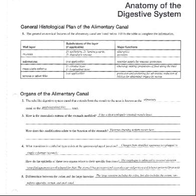
December 2019 27
Breskvar V Wall - Uni Study Guides 1k6t4f
December 2019 45