Membrane Physiology 3w2a3j
This document was ed by and they confirmed that they have the permission to share it. If you are author or own the copyright of this book, please report to us by using this report form. Report 2z6p3t
Overview 5o1f4z
& View Membrane Physiology as PDF for free.
More details 6z3438
- Words: 7,949
- Pages: 223
MEMBRANE PHYSIOLOGY
1
Objectives
• At the end of this course students are expected to: Write components of cell membrane and their functions Understand cell signaling by electrical excitation Define what membrane potential mean List different ion channels and their functions 2
Cont’d Differentiate action potential and graded potential Define synapse and its function Compare and contrast electrical and chemical synapses Discuss cell communication Explain cellular transduction process 3
what is cell? • The cell is the fundamental structural and functional working unit of all organisms • It possesses and exhibits the basic properties of living matter. • Cell is the smallest living unit and it is microscopic. • Cells are structural & functional unit in our body. • Human body is assembled of eukaryotic cells. • There are 75-100trillion cells in our body 4
• The three fundamental cell theories are:• 1. Cells come from pre-existing cells. • 2. A cell is a fundamental unit of life. (i.e. all living things are made up of cells.) • 3. Cell is capable of maintaining its existence (metabolize, grow & reproduce on its own).
5
• Basic cell functions:– Obtaining food (nutrients) & O2 from nearby env’t . – Performing various metabolic energy production – Elimination of waste products (CO2 & others) – Synthesis of materials for cellular structure, growth,& carrying out cellular functions – Sensitivity & responsiveness to changes in its env’t – Control of material exchanges b/n cell & env’t. – Moving materials within the cell in carrying out cellular activities. 6
Eukaryotic Cell Structure and function 3 main parts of the cells are: –
Nucleus the largest organelle in the cell
–
Cytoplasm -the region b/n the plasma membrane & nucleus.
–
•
Filled with intracellular fluid (cytosol).
•
Most organelles are suspended in cytoplasm Plasma membrane- the outer covering of the cells. 7
Cell structure and function
8
•
Cytoplasm
Material enclosed by plasma membrane
• Is a watery solution of minerals, gases, organic molecules, and cell organelles that is found between the cell membrane and the nucleus. • It is a fluid matrix, the cytosol, which consists of 80% to 90% water, salts, organic molecules and many enzymes that catalyze reactions. • Cytosol is the water portion of cytoplasm, and many chemical reactions take place within it.
9
Cell Organelles • Are intracellular structures, often bounded by their own membranes, that have specific functions in cellular metabolism • Highly organized structures – are basic machinery found within cells. – are responsible for the functioning of the cell. – perform essential activities for survival of cell. 10
Nucleus
11
• Endoplasmic Reticulum (ER) -Network of membranes made up of tubes, sacs & chambers called cisternae • Attached to the nuclear envelope -2 forms:
12
Cont’d a. Rough endoplasmic reticulum (RER) -studded with ribosomes -ribosomes synthesize proteins and feed them into RER cisternae to be modified(e.g. +carbs = glycoprotein) -modified proteins put into transport vesicles to go to Golgi -these proteins for exocytosis or use in membrane b. Smooth endoplasmic reticulum (SER) -tubular membranes -no ribosomes 13
Cont’d Functions of SER: 1. lipid metabolism (synthesis, breakdown,transport) 2. synthesis of steroid hormones 3. detoxification of drugs 4. breakdown of glycogen to glucose 5. store ions (e.g. Ca2+)
14
• Endoplasmic reticulum.
15
Golgi apparatus – Is a set of stacked membranes – near nucleus but not attached – functions to modify, concentrate, & sort export proteins – It modify proteins to the final stage of production – adds sugar molecules to side groups, protects proteins from breakdown – Packages proteins into vesicles for secretion or internal use 16
Cont’d
17
18
Cytoskeleton –structure that gives mechanical to the cell • helps cell to maintain its shape
• Enables a cell to change shape in an adaptive manner • help for cell motility: – organelle movement, – muscle contraction, and
• Plays a regulatory role by transmitting signals from cell's surface to its interior.
19
• cytoskeleton
20
Plasma Membrane
21
Cell membrane/plasma membrane •Is very thin structure that covers the outer surface of a cell with about 7-10 nm thick. • It separate cellular content from environment(It forms boundary of the cell). • It control movement of material into and out of the cell. •It is lipid bilayer (made of phospholipids arranged in double layer). •The phospholipids has both hydrophobic and hydrophilic regions.
22
Cont’d
• Thus phospholipids in cell membrane are held together by hydrophobic interactions • The lipid layer in the middle of the membrane is impermeable to the usual water-soluble substances, such as ions, glucose, and urea. • Conversely, fat-soluble substances, such as oxygen, carbon dioxide, and alcohol, can penetrate this portion of the membrane with ease
23
• The phospholipids in the cell form a bilayer that acts as a semi permeable boundary between the cell membrane and its external environment •
It is composed almost entirely of proteins and lipids
• Membrane composition is 55% proteins; 25% phospholipids; 13% cholesterol; 4% other lipids and 3% carbohydrates.
24
Membrane Functions • Protection( & retain the cytoplasm) • Cell to cell communication(Communication via receptors) • Selectively permeable - controls what goes in and out of cell or organelle • Respond to environment • Recognition • Signal transduction interface
25
Components of the cell membrane • Contains lipids, proteins and carbohydrates – Lipids • Phospholipids -self assemble into bilayer • Cholesterol-resist osmotic lysis – Proteins • Integral -span width of membrane • Peripheral-adhere to inner or outer surface – Carbohydrates linked to other molecules as glycolipids and glycoproteins 26
27
Cont’d
28
Cell Membrane Structure 1. Lipids • Phospholipids: – Hydrophilic head (likes water) – Hydrophobic tails (cannot be in with water) • Cholesterol: o Stiffens membranes o Can move in any dimension through membrane o contributes to both the fluidity and the stability of the membrane. 29
30
•Cholesterol molecules are tucked in between the phospholipids molecules, where they prevent the fatty acid chains from packing together and crystallizing, •Through their spatial relationship with phospholipid molecules, cholesterol molecules also help stabilize the phospholipids’ position 31
Cont’d
The lipid bilayer serves three important functions: 1. It forms the basic structure of the membrane. –
The phospholipids can be visualized as the “pickets” that form the “fence "around the cell.
2. Its hydrophobic interior serves as a barrier to age of water-soluble substances between the ICF and ECF. 3. It is responsible for the fluidity of the membrane
32
2. Proteins: ½ mass of membrane a. Peripheral proteins: - Adhere to inner or outer surface –
Hydrophilic and readily dissociated from membrane.
–
Free, floating on the surface (stud the inside and the outside of the membrane) and for about 30% of the membrane proteins.
–
Bind specific hormones and proteins on cell membrane.
–
They also function as enzymes. 33
34
b. Integral proteins • span width of membrane •
Transmembrane proteins (trans means “across”).
• Exist as separate globular units running through the width of the cell membrane • Partly hydrophilic (polar and protruding to cell surface) and partly hydrophobic (non-polar and embedded in the lipid bilayer). • Protruding part may often carry CHO chains or lipids attached to their tips like flags .
35
Cont’d • Tightly associated with membrane and for about 70% of the membrane proteins. •
Many of them provide structural channels (or pores) through which water molecules and water-soluble substances, especially ions can diffuse in and out of the cell.
•
Other integral proteins act as channels, carriers, pumps, enzymes and receptors. 36
Cont’d
37
Function of cell membrane proteins • • • • • • •
Transporter (Channels + Carriers) Receptors Enzymes Signal Transducers Cell-to-cell recognition Cell-to-cell junction 38
3. Carbohydrates • Polysaccharides…Hydrophilic • Always found on the exterior surface of cells. • Glycocalyx: – Glycoproteins = CHO + Proteins – Glycolipids = CHO + Lipids Used for cell -to-cell recognition • These carbohydrate chains play an important role in recognition of “self ” and in cell-to-cell interactions. • Cells can recognize other cells of the same type and to form tissues 39
40
Cellular Transport Function/ Membrane transport • Transport across cell membrane – ive transport and – Active transport • ive transport – Do not require energy – From high to low concentration – Down concentration gradient • Eg. Osmosis • Simple diffusion • Facilitated diffusion • Osmosis is net flow of water molecule from high water content to low water content across selectively permeable membrane. 41
ive Transport Mechanisms cont’d Cell uses no energy Molecules move randomly from an area of high concentration to an area of low concentration (HighLow). At equilibrium, movement is equal in both directions Three types: 1. Simple Diffusion: .Free movement of a substance from one part of a solution to another occurring in both directions. .Net movement occurs from an area of high concentration to another area of low concentration.
42
Rate of diffusion is affected by: – Viscosity of solution – Molecular weight – Concentration gradient: Difference in concentration between two points – Permeability of the membrane / medium – Available surface area and thickness of membrane – Distance traveled across which diffusion must occur (inverse) – Solvent state (gas > liquid > semisolid)
43
2. Facilitated Diffusion -Require no energy -Is carrier mediated transport - Cellular Transport From a-
Glucose molecules
High
High Concentration Cell Membrane Low Concentration Through a Transport Protein
L
Protein channel
uses a carrier to facilitate (assist) the transfer of a particular substance across the membrane “downhill” from high to low concentration .
Go to Section:
44
Facilitated diffusion includes cont’d i. carrier-mediated transport- uniport and symport ii. carrier-mediated exchange- anti-port iii. ion channels and channels for other small molecules
Uniport
Classes of carrier proteins
A
Symport A
B
Antiport A
B 45
3. Osmosis • the diffusion of water across a semi-permeable membrane • Water moves from solution with lower concentration of osmotically active solutes • Water moves from dilute solution to concentrated solution • Osmotic potential is the total of all dissolved particles
46
Active Transport o Active transport requires Energy. o Movement from an area of low concentration to an area of high concentration (Low High). o When cells must move materials in an opposite direction against a concentration gradient. - Proteins or Pumps are found in the cell membrane transport molecules across the membrane. o Three types 1.Protein Pumps 2.Endocytosis 3.Exocytosis 47
Cont’d
48
Cont’d Active transport •
Requires the carrier to expend energy to transfer its enger “uphill” against a concentration gradient, from an area of lower concentration to an area of higher concentration
• Active transport is carrier-mediated transport that uses energy and moves a substance against its concentration gradient
49
Cont’d Active transport comes in two forms.
In primary active transport, • Energy is directly required to move a substance against its concentration gradient • The carrier splits ATP to power the transport process
50
Cont’d • The most important example is a sequentially active Na–K ATPase pump (Na–K pump for short) found in the plasma membrane of all cells. • This carrier transports Na out of the cell, concentrating it in the ECF, and picks up K from the outside, concentrating it in the ICF . • The pump has high affinity for Na on the ICF side
51
Cont’d
52
Cont’d
• In secondary active transport, – energy is required in the entire process, but it is not directly used to produce uphill movement. – the carrier does not split ATP; instead, it moves a molecule uphill by using “secondhand” energy stored in the form of an ion concentration gradient (most commonly a Na gradient). – This ion gradient is built up by primary active transport of the ion by a different carrier. 53
The Na–K pump plays three important roles: 1. It establishes Na and K concentration gradients across the plasma membrane of all cells; • these gradients are critically important in the ability of nerve and muscle cells to generate electrical signals essential to their functioning 2. It helps regulate cell volume by controlling the concentrations • of solutes inside the cell and thus minimizing osmotic effects that would induce swelling or shrinking of the cell. 3. The energy used to run the Na–K pump also indirectly • serves as the energy source for secondary active transport
54
Membrane transport of macromolecules & particles • These large particles are transferred between the ICF and ECF • by being wrapped in a membrane-enclosed vesicle, a process known as vesicular transport. Vesicular transport – requires energy expenditure by the cell, – Is an active method of membrane transport. – Energy is needed to accomplish vesicle formation and vesicle movement within the cell.
• Transport into the cell in this manner is termed endocytosis, • whereas transport out of the cell is called exocytosis 55
• Endocytosis - cell takes in large particles by engulfing them 1. Phagocytosis - "cell eating" - extensions off cytoplasm surround a particle and package it within a food vacuole and then the cell engulfs it. Ex. Amoebas use this process. 2. Pinocytosis – “cell drinking” the process of taking up liquid from the surrounding environment. Tiny pockets form along the membrane, fill with liquid, and pinch off.
56
Exocytosis • cell gets rid of particles, opposite of endocytosis • the vesicles created by the Golgi Body. • These are removed from the cell by exocytosis.
57
Cont’d
58
Cell signaling(control of cell function) Terminology Ion Atom / molecule that have an electrical charge.
Anion Negatively charged ion (e.g., Cl −).
Cation Positively charged ion (e.g., Na+, K+, Ca2+).
Influx of ions Flow of ions into the cell (ECF to ICF).
Efflux of ions Flow of ions out of the cell (ICF to ECF). 59
Cell signaling by electrical excitation
Membrane Potential • Electrical energy difference between the inside and outside of the cell. • Separation of opposite charges across the plasma membrane • Thus ICE is relatively -ve while ECE is relatively +ve
Em = Vin – Vout, where Vin = Potential on the inside of the cell Vout = Potential on the outside Em = Membrane potential (mV) 60
• All cells have membrane potentials. • Membrane potential charge separation across the membrane. • Any change of a membrane’s permeability of ions causes a change in Em.
61
Resting Membrane Potential • Steady transmembrane potential of a cell that is not producing an electrical signal. • No net flow of ions across the plasma membrane. (No net inward current) • Always negative in nerve and muscle cells. • Magnitude is κ+ for individual cell types. o Nerve, cardiac and skeletal muscle: -55 to -90mV o Smooth muscle: -55 to -30mV
62
• Inexcitable cells have RMP. • RMP is necessary for the cell to fire an action potential. • Resting membrane potential is nearly equals to that of EK
NB. Excitable cells (nerve cells and muscle cell) have the ability to produce rapid, transient changes in their membrane potential when excited.
63
64
65
Determinants: Origins of Em ive determinants Active determinant ive Determinants A. Biochemical nature of plasma membrane of the cell. Lipid bilayer (7nm = 60Å): Selective permeability • Extracellular: • Intracellular:
+VE - VE
66
B. Asymmetrical/Unequal distribution of ions across the membrane
67
Concentration of ions Inside Sodium (Na+)
12mM
Outside 145mM
Electrostatic [ ] gradient
pressure
into cell
into cell
Potassium (K+) 150mM
5mM
out of cell
Chloride (Cl-)
125mM
in
out
2.5mM
into cell
into cell
9mM
Calcium (Ca2+) 10-4mM Organic anions:
into cell
Fixed anions (Proteins, nucleotides, polyphosphates…)
68
C. Leakage (Leak, non-gated, ive, resting) channels • Leak K+ channels, leak Na+ channels, leak Cl- channels • Leakage K+ channels are open at resting potential more than Na+, Cl-
• Resting membrane potential is nearly equals to that of the EK.
69
D. Diffusional force
70
Nernst Equation/Nernst Potential •Used to calculate the equilibrium potential for a given ion with differing concentrations across a membrane •Equilibrium potential is a measure of membrane potential i. Conc. of the ion inside and outside ii. Temperature of the solution iii. The valence of the ion iv. The amount of work required to separate a given quantity of charge. Ex = RT ln [X]o (mV) zF
[X]I
71
Where R = gas constant = 8.315jk-1mol-1 T = TKelvin = 271.16 + TCelcius F = z = [X]o [X]i
NB.
Faraday's constant = 96, 485 Cmol-1 Valence of the ion (+1, -1, +2) = Concentration of the ion outside the cell = Concentration of the ion inside the cell
Ex = 61 log10 [X]o ── [X]i Z Nernst Equation predicts Equilibrium Potential (Eion). 72
Goldman-Hodgkin-Katz Equation •Used to calculate membrane potential •Considers the relative permeabilities and concentration gradients of all permeable ions
Em = RT ln PK[K+]o + PNa[Na+]o +PCl[Cl-]i F PK[K+]i + PNa [Na+]i + PCl[Cl-]o
(Permeability = PK: PNa: PCl, 1.00: 0.04: 0.45)
73
Goldman-Hodgkin-Katz Equation 1. The contribution of any ion to the Em is determined by
the membrane’s permeability to that particular ion. 2. The higher the permeability of the membrane to one ion relative to the others, the more that ion will contribute to the Em. 3. The contribution of the impermeable ion to the Em will be zero. NB. GHK = predicts Resting Membrane of a cell. 74
Active Determinant Na+-K+-ATPase (Na+-K+ pump) A carrier molecule uses the membrane-bound ATPase. Primary active transport process (consumes ATP, pumps against conc. or electrical gradient). Operates as antiporter (coupled transporter): Pumping 3Na+ out of the cell Pumping 2K+ in . 75
Functions i. Maintenance of gradient of Na+ and K+ across the cell membrane •Controls cell volume ( Na+ regulating osmotic forces) ii. Control of membrane potential and excitability.
Na+–K+–ATPase 76
Na+–K+–ATPase 77
Ion channels • Electrical signals are produced by changes in ion movement across the plasma membrane.
• The water-soluble ions responsible for carrying charge cannot penetrate the plasma membrane’s lipid bilayer,
• these charges can cross the membrane only through channels specific for them or by carrier-mediated transport.
• Membrane channels may be either leak channels or gated channels 78
leak channels • which are open all the time • permit unregulated leakage of their specific ion across the membrane through the channels.
Gated channels
• Are complexes membrane protein. • These channels are normally closed, – but open in response to appropriate signal (stimulus) – Then allow specific ions to flow into or out of the cell 79
Gated channels
cont’d
• Have gates that can be open or closed, permitting ion age through the channels when open and preventing ion age through the channels when closed. • Gate opening and closing results from a change in the conformation (shape) of the protein that forms the gated channel. 80
• There are four kinds of gated channels, – depending on the factor that causes the change in channel conformation:
(1) voltage-gated channels (.These channels are regulated by the membrane potential. (.open or close in response to changes in membrane potential; (•) The most prominent of these channels is the voltage-gated Na+ channel. (•) Its opening underlies the initiation of an action potential
81
Cont’d (2) chemically gated channels/ Ligand-gated ion channels: •
change conformation in response to binding of a specific extracellular chemical messenger to a surface membrane receptor;
• Are regulated by a specific molecule that binds to the ion
channel.
• Postsynaptic neurotransmitter receptors are ligand-gated ion
channels 82
Cont’d (3) mechanically gated ion channels • respond to stretching or other mechanical deformation – Touch receptors in the skin and – Receptor cells in the inner ear are examples of mechanically gated ion channels.
• These channels open through the mechanical deflection that pries or pulls the channel open (4) thermally gated channels • respond to local changes in temperature (heat or cold). • The channel protein acts as a thermometer, and a change in temperature opens the channel pore 83
84
Cont’d ive Ion Channels • Found on dendrites, cell body, and axon.
Chemically-gated Ion Channels • Found on dendrites & cell body.
Voltage-gated Ion Channels • Found on axon hillock, unmyelinated axons and at nodes of Ranvier on myelinated axons. 85
Cont’d
86
• ΔEm: initiate electrical signaling in excitable cells • Basis of signaling in the NS. • Types: 1. Graded potentials 2. Action potentials
87
1.Graded potentials a. Def.:-
local changes in membrane potential (Em) in
either depolarizing or hyperpolarizing direction. •.Are local changes in membrane potential that occur in varying grades or degrees of magnitude or strength. •.which serve as short-distance signals
88
Graded potential cont’d • It is local electrical disturbance • Propagate like wave in all direction • Its propagation is decreasing (as it go away from point of origin its amplitude ) • Its amplitude depend on strength of stimulus • Strong stimulus generated large graded potential that can propagate longer distance • Occurs in dendrite, cell body or motor end plate – b/c these parts lacks voltage gated ion channel
• If it reaches threshold potential it generate action potential. 89
• Sub-threshold stimulus do not generate action potential (weak stimulus < threshold not generate action potential). • Threshold is minimum required potential to generate action potential
90
• Weak stimulus cause opening of few ion channels. • Influx of few ions- little disturbance- easily disappear-with out causing action potential. • Strong stimulus cause opening of many ion channels. • Influx of many ions- large disturbance-not easily disappear-propagate longer distance-if it reaches threshold point initiate action potential. 91
Subthreshold and suprathreshold graded potentials in a neuron. In (a), the graded potential is below the threshold value (T) of *55 mV as it reaches the trigger zone. In (b), the graded potential is above the threshold value when it reaches the trigger zone. 92
Summary of GP Features: 1. Depolarizing or hyperpolarizing 2. Variable in amplitude and duration (AM) 3. Conducted decrementally 4. Can be summed 5. Has no threshold 6. Has no refractory period
93
Types:
Receptor Potentials/Generator Potentials
Graded Potentials
Pacemaker Potentials Excitatory Postsynaptic Potentials/EPSP
Synaptic Potentials
Inhibitory Postsynaptic Potentials/IPSP
94
Mechanism of signaling:
Stimulus (physical, mechanical, chemical, electrical…) Sensory receptors Transform stimulus energy/Transduction Ion channels open Inward flow of current Depolarization ΔEm Receptor potential/Generator potential Action Potential CNS ...→ RESPONSE. 95
Action Potentials Def: rapid, transient, self-propagating electrical excitation in the plasma membrane of excitable cells. Genesis/Initiation: electrical perturbation ΔEm AP. Sequential opening of voltage-gated channels of Na+ and K+. which signal over long distances
96
Part I
Characteristics of AP: 1. All-or-none phenomenon. •Always be full sized. •It will not get lost along the way. •Rapid and reliable information.
2. Has threshold. 3. Amplitude and duration is κ 4. Always depolarizing. 5. Has refractory period. 6. Nondecremental.
97
Part I
98
Cont’d
All or none law The principle that the strength by which a nerve or muscle fiber responds to a stimulus is not dependent on the strength of the stimulus If the stimulus is any strength above threshold, the nerve or muscle fiber will give a complete response or otherwise no response at all
99
100
cont’d Physiological basis of AP When the threshold level is reached Voltage-gated Na+ channels open up Since Na conc. outside is more than the inside Na influx will occur Positive ion coming inside increases the positivity of the membrane potential and causes depolarization When it reaches +30, Na+ channels closes Then Voltage-gated K+ channels open up K+ efflux occurs Positive ion leaving the inside causes more negativity inside the membrane Repolarization occurs 101
Cont’d Since Na+ has come in and K+ has gone out
Membrane has become negative But ionic distribution has become unequal Na+/K+ pump restores Na+ and K+ conc. By pumping 3 Na+ ions outward and 2+ K ions inward
102
• Stages of Action Potential. Stages of AP are Resting stage Depolarization stage Repolariztion stage Hyperpolarization stage
103
• Events in different stages of action potential.
104
Phases of an AP and Inward and outward currents 105
Phases and Ionic Basis of Action Potential 1.Threshold Potential Minimum value of Em at which an action potential will occur . Em is reduced to a critical level excitable cells undergo rapid depolarization. • Initiated by rapid opening of fast Na+ channels. AP occurs only when the NET movement of positive charge is inward (gNa+ > gK+ or Na+ influx > K+ efflux).
106
Magnitude is 10-20mV (-70mV -55mV). Strength of stimuli • Subthreshold • Threshold Stimuli
107
• Suprathreshold stimuli
108
2. Depolarization Phase/ upstroke • ↑gNa+ flow of Na+ into the cell • m, h gates opened.
109
Sodium channels exert positive on membrane potential. 110
3. Overshoot/peak of the action potential i. Portion of the AP during which the membrane is positive. ii. Magnitude: 30 to 40mV iii. Approaching ENa+
Potassium .
channels
exert
negative
111
4. Repolarization/Downstroke i. Rapid return of the membrane towards its RMP.
ii. Reasons: a. Time-limited nature of Na+ permeability (gNa+ is short-lived) b. Closure of h-gate of Na+ Na+ inactivation c. ↑gK+ (delayed opening of K+ channels, n-gates open, activated) 112
5. Afterpotentials/undershoot •Membrane becomes more negative than its RMP at the end of the action potential. 5.1. Hyperpolarization afterpotential (-70mV -72/-73mV, 40ms) n gates opened ↑K+ out of the cell ↑ inside negativity excitability.
113
Refractoriness/Refractory period i. Def. an interval during which it is more difficult to elicit an AP. (∵ voltage and time-dependent nature of gating particles) . ii. Types: a. Absolute refractory period b. Relative refractory period
114
a. Absolute Refractory Period: •Another AP can not be elicited, regardless of the strength of the stimulus. •Begins at the start of the upstroke and extends into the downstroke.
•During the upstroke m gates are opening. •During the early portion of the downstroke Na+ channels are inactivated by h gates. •Ends when the number of inactivated Na+ channels is reduced by repolarization. NB. ARP Ensures ONLY one-way of propagation of APs along an axon. 115
b. Relative Refractory Period • A second AP can be elicited if the stimulus is adequate. • Stimulus must be greater than normal (suprathreshold) i. The Na+ channels are still inactivated. ii. More K+ channels than normal are still open. NB. RRP helps to limit frequency of AP
116
Cont’d
Functions of refractory period Ensures ONLY one-way of propagation of APs along an axon. Imposes a limit on the maximum rate a neuron can fire. Prevents APs from summating.
117
Refractory periods 118
119
120
Na+ channel: 2 gating particles a. m-gate: covers the EC side of the Na+ channel. b. h-gate: covers the IC side of the Na+ channel. • m-gate open: activation of Na+ channel • h-gate close: inactivation of Na+ channel c. Both m and h gates must be open for Na+ to flow thru the Na+ channel.
121
122
Conformations of voltage-gated sodium channels
cont’d
Voltage-gated Na+ channel can exist in three conformations:• Closed but capable of opening (activation gate closed, inactivation gate open)
• Activated, or open (both gates open). • Inactivated, closed and not capable of opening (activation gate open, inactivation gate closed)
123
Models of Gating Particles of Voltage-gated Na+ channel… 124
K+ channel: ONE gating particle: n-gate • n-gate covers the IC side of the K+ channel • n-gate must be open for K+ to flow thru the K+ channel. n-gate open: activation of the K+ channel K+ channel does not have an inactivation gate.
125
Conformations of voltage-gated potassium channel
126
Propagation of Action Potential Types: i. Cable conduction/Continuous/Sweeping/conduction •
Unmyelinated nerve
•Speed of AP √diameter of the fiber (√1μm = 1m/s = slow)
127
Cable Conduction 128
ii. Saltatory Conduction • Occurs in myelinated nerve 1. Myelin sheath: covers some axon is made from cytoplasm of glial cells: Schwann cells PNS Oligodendroglia CNS. 2. Nodes of Ranvier - little gaps in the myelin sheath The electrical signal is said to jump from node to node, thus it is called saltatory conduction Significance: i. Myelin sheath: high resistance + lower capacitance. ii. increase velocity (fast, unidirectional)
129
130
131
Saltatory conduction 132
Saltatory conduction 133
Saltatory conduction 134
135
Factors affecting propagation of AP a. Type of fiber (myelinated or non myelinated neuron) •Velocity myelination Velocity in myelinated n > nonmyelinated nerve b. Geometry of the fiber: diameter of axon Larger axon conduct signal faster than smaller axon Because there is less friction between the moving charged particles (Na+ and K+) and sides of axon in larger axon • Velocity diameter of the fiber (↑SA, ↓R)
c. Temperatureincrease temp. chemical reaction speeds up ↓o C ↓ rate of change of permeability (effect on the enzymatic machinery)
136
Cont’d
137
Transmission of excitation from cell-to-cell (synapses) Synaptic transmission • Is the transfer of signals from one cell to another. •Is communication among neurons, with muscles and glands •A neuron may terminate on one of three structures: –a muscle, a gland, or another neuron. •Therefore, depending on where a neuron terminates, it can cause –a muscle cell to contract, –a gland cell to secrete –another neuron to convey an electrical message along a nerve pathway, or some other function.
138
Cont’d Synapse A site at which an impulse is transmitted from one cell to another A special zone of at which one neuron communicates
with another Is the functional connection between a neuron and a second cell. In the CNS, this other cell is also a neuron. In the PNS, the other cell may be either a neuron or an effector
cell within a muscle or gland
139
Cont’d • Neuron-neuron synapses usually involve a connection between the axon of one neuron and the dendrites, cell body, or axon of a second neuron. • These are called, respectively axodendritic, axosomatic, and axoaxonic synapses. •
In almost all synapses, transmission is in one direction only—from the axon of the first (or presynaptic) neuron to the second (or postsynaptic) neuron.
•
Most commonly, the synapse occurs between the axon of the presynaptic neuron and the dendrites or cell body of the postsynaptic neuron.
140
Cont’d
141
Cont’d
Synapses are typically junctions between presynaptic and postsynaptic neurons. • There are two types of synapses: – Electrical synapses and – Chemical synapses, depending on how information is transferred between the two neurons. 142
Chemical synaptic transmission • There is no continuity between the cytoplasm of the presynaptic terminal and postsynaptic neuron. • The cells are separated by synaptic clefts, which are fluid-filled gaps (about 20–50 nm). • The presynaptic and postsynaptic membranes adhere to each other by a matrix of extracellular fibrous protein in the synaptic cleft. • Communication is achieved via neurotransmitters 143
Cont’d
144
Structural components of a chemical synapse i. Presynaptic Terminal: Neurotransmitter(NT)synthesizing transporters, reuptake Transporters …
enzymes,
synaptic
Vesicles • Size a. Small clear: Ach, glycine, GABA, glutamate b. Small dense core vesicles: Catecholamines c. Large dense core vesicles: Neuropeptides • Shape: Round vesicles: excitatory transmitters Flattened vesicles:inhibitory transmitters 145
vesicle
Synaptic transmitters
Neurotransmitters: •
Are molecules released by presynaptic neurons and are the means of communication at a chemical synapse.
•
Bind to neurotransmitter receptors, which can be coupled with – an ion channel (ionotropic receptors) or with – an intracellular signaling process (metabotropic receptors).
•
Neurotransmitters are specific for the receptor they bind to and elicit a specific response in the postsynaptic neurons, resulting in either an excitatory or inhibitory signal
146
Active zone: is a site of neurotransmitter release •Voltage-gated Ca2+ channels responsible for rapid NT release. ii. Synaptic cleft: • Width: 30nm (x = 20-50nm) • Contents: Basal lamina Inactivating enzymatic system iii. Postsynaptic terminal: •Transmitter-gated ion channels (ligand-gated ion channels/Ionotropic receptors)
•G-Protein coupled receptors, Gprotein-gated ion channels/Metabotropic receptors •2nd messenger cascades (cAMP, cGMP, PLC…) 147
Neurotransmitter receptors are:
cont’d
i. Ionotropic: fast, open ionic gates and allow the flow of current thru the postsynaptic membrane (eg., Ach).
Ionotropic Receptors Ligand-gated Ion Channels Directly-gated Ion Channels 148
149
ii. Metabotropic: slower, longer lasting change, affecting cellular permeability.. (using G Proteins, Ca2+, calmodulin, cAMP...) (eg., NE).
Metabotropic Receptors G Protein-gated Ion Channels Indirectly-gated Ion Channels 150
151
152
153
154
Types of chemical synapses (On morphological basis…) a.
Excitatory • • • • •
b.
Round synaptic vesicles Wide synaptic cleft Large active zone Asymmetric postsynaptic density Axodendritic synapse
Inhibitory • • • • •
Flattened synaptic vesicles Narrow synaptic cleft Small active zone Symmetric postsynaptic membrane Axosomatic synapse
155
Cont’d
Excitatory synapse Presynaptic neuron neurotransmitter (Ach, glutamate, serotonin ...) open cation channels influx of Na+ depolarization of the postsynaptic membrane towards the threshold potential excitatory post synaptic potential(EPSP). • Neuron →
action potential
• Muscle → contraction • Glands
→
secretion
156
The generation of an EPSP 157
Characteristics of excitatory postsynaptic potentials(EPSPs) • Transient depolarization. •
Excitatory because Em moves closer to threshold.
•
Increase in gNa+ .
•
Na+ influx causes depolarization
158
Inhibitory synapse Presynaptic neuron neurotransmitters (GABA, glycine ...) open Clchannels Cl- enters into the cell postsynaptic membrane hyperpolarized suppress firing in postsynaptic cell IPSP.
The generation of an IPSP 159
Cont’d
160
Characteristics of IPSPs • Transient hyperploarizations. • Inhibitory because Em moves further away from its threshold. • Increased conductance to Cl• Cl- influx causes hyperpolarization. • Can be produced by increased gK+ + accelerated K+ efflux or closure of Na+ and Ca2+ channels.
161
Sequence of events at chemical synapses: Action potential in presynaptic cell ↓ Depolarization of plasma membrane of the presynaptic axon terminal ↓ Entry of Ca2+ into presynaptic terminal ↓ Release of the transmitter by the presynaptic terminal ↓ Chemical combination of the transmitter with specific receptors in the plasma membrane of the postsynaptic cell ↓ Transient change in the conductance of the postsynaptic plasma membrane to specific ions.
↓ Transient change in the Em of the postsynaptic cell 162
Sequence of events at chemical synapses 163
Sequence of events at chemical synapses
164
165
Integration of synaptic events/ Synaptic Integration
166
Synaptic Integration 1. A central neuron receives both excitatory and inhibitory signals. 2. Excitatory and inhibitory signals are integrated into a single response by the postsynaptic cell. 3. Excitatory synaptic action is mediated by glutamate-gated channels... that conduct Na+ and K+. 4. Inhibitory synaptic action is usually mediated by GABA- & glycinegated channels that conduct Cl-. 167
5. Net effect of inputs depends on the location/proximity, size, and shape of the synapse: • • •
Synapses on cell bodies: inhibitory Synapses on dendritic spines: excitatory. Synapses on axon terminals: modulatory.
6. Net effect is sum of excitatory + inhibitory signal inputs.
168
Synaptic potentials • EPSPs and IPSPs are graded potentials. • Graded potentials can be of varying magnitude, have no refractory period, and can be summed (added on top of one another). • The postsynaptic neuron can be brought to threshold by either – temporal summation or – spatial summation.
169
Synaptic Integration: Summation •
Temporal
•
Spatial
170
Temporal summation 171
Spatial summation 172
Spatial summation
173
If excitatory signals > inhibitory signals →depolarization/excitatory.
174
If inhibitory signals > excitatory signals → hyperpolarization/inhibitory.
175
Action potential is initiated at the initial segment, axon hillock.
176
EPSP + IPSP = GPSP
177
178
179
Electrical synaptic transmission
• Electrical Synapses (Ephatic or electro tonic transmission) • In ES ion channels connect the cytoplasm of the presynaptic and postsynaptic cells • Two neurons can be coupled electrically to each other via gap junctions • Rapid electrical signaling and information. • (In reflex reactions: escape and defensive responses)
180
cont’d Found in neuronal + non-neuronal cells. • Neuroendocrine cells of hypothalamus (Magnocellular + Parvocellualr cells) • Lateral vestibular nucleus, inferior olive, cerebellum, neocortex • Thalamus, striatum, hippocampus, retina, olfactory bulb • Neuroglial cells (astrocytes, Schwann cells) • Myocardial cells • Smooth muscle cells • Epithelial cells + hepatocytes • Lens
Do not generally allow inhibitory actions or long-lasting changes in effectiveness. 181
A gap junction is a protein pore complex (connexon) that lets ions and other small molecules move between cells . 182
Characteristics of electrical synapses A ΔEm in one cell is transmitted to the other cell by the direct flow of current (cytoplasmic bridge/gap junction between cells). No synaptic delay (direct interactions between neighboring cells). Allow conduction in both directions(information flow is bidirectional). Significance: i. Intracellular signaling during embryonic development. ii. Synchronization of impulses. 183
Cont’d
184
Cell communication by Autacoids and Paracrine hormones • Communication between cells is largely orchestrated by extracellular chemical messengers. • Intercellular communication can take place either • directly or • indirectly • Direct intercellular communication involves physical between the interacting cells • Through gap junctions • Through transient direct linkup of surface markers 185
Cont’d Gap junctions.
• Is the most close means of intercellular communication
• the minute channels that bridge the cytoplasm of neighboring cells in some types of tissues.
• Through gap junctions, small ions and molecules are directly exchanged between interacting cells without ever entering the extracellular fluid. 186
Cont’d Transient direct linkup of surface markers. •
Some cells, such as those of the immune system, have specialized markers on the surface membrane,
•
that allow them to directly link with certain other cells that have compatible markers for transient interactions.
• This is how the phagocytes of the body’s defense system specifically recognize and • selectively destroy only undesirable cells, such as cancer cells, while leaving the body’s own healthy cells alone.
187
Cont’d
188
Indirect communications • The most common means by which cells communicate with one another is indirectly through extracellular chemical messengers or signal molecules, of which there are four types:
– Paracrines/ autocrines – neurotransmitters, – hormones, and – neurohormones. • In each case, a specific chemical messenger, the signal molecule, is synthesized by specialized controlling cells to serve a designated purpose 189
1. Paracrine signaling • Paracrines are secreted by cells into the extracellular fluid and affect neighboring cells of a different type(Local chemical messengers ) • They are secreted into the ECF and diffuse to adjacent cells. • Distributed by simple diffusion within interstitial fluid, • Their action is restricted to short distances.
190
Cont’d
2. Autocrine signaling • Autocrines are secreted by cells into the extracellular fluid and affect the function of the same cells that produced them by binding to cell surface receptors • Autocrine action: the hormone acts on the same cell that produced it. 191
3. Endocrine signaling • Hormones are long-range chemical messengers • Secreted into blood by endocrine glands in response to signal. • Exert effect on target cells some distance away from release site. • To respond to a chemical signal, a target cell must have a receptor protein for it. • Hormones reach and bind to receptors of target cells via circulating blood. 192
4. Neurohormones • are hormones released into the blood by neurosecretory neurons. • The neurohormones are then distributed through the blood to distant target cells. • An example is ADH, a hormone produced by nerve cells in brain that promotes water conservation by kidneys during urine formation. 193
Autacoids (Local Hormones)
-- Autocrine secretions act on the cells which secrete them -- Paracrine secretions, secreted by some cells and act locally. 194
Cont’d
195
Cont’d
Autacoids - local hormones • Are biological factors which act like local hormones, have a brief duration, and act near the site of synthesis • Thus the word autacoids was used for substances that act within restricted, local areas near their site of synthesis,
• These substances usually have a brief lifetime. 196
cont’d • Heterogeneous substances have been included as autacoids. These are: •
•
•
Amine Derived – Histamine – 5 Hydroxytryptamine (serotonin) Lipid derived – Prostanoids (prostaglandins, thromboxanes) – Leukotrienes – Platelet Derived Factor Peptide – Angiotensin – Kinins (Bradykinin, kallidin) – Vasoactive intestinal Peptide – Neurotensin – Substance P – Calcitonin Gene related Peptide
197
Histamine : is a bioactive amine , widely distributed in animal and plant tissues. • In human body, histamine present in skin , GIT , lungs , CNS , CVS, basophiles. • The histamine acts as a Paracrine signal, diffusing to capillaries in the immediate area of the injury and making them more permeable to white blood cells and antibodies in the plasma.
198
Physiological role of histamine: 1- neurotransmitter in CNS. 2- Micro-circulatory regulation. 3- control sleep and alertness . 4- correlates with fetal development . 5- wounds healing. 6- Has a role in thermal and body weight regulation. 7- Gastric acid secretion.
199
Serotonin: is one of most important autacoids • Serotonin is a bioactive amine that can be synthesized by hydroxylation of amino acid tryptophan to 5-hydroxy tryptophan ( by enzyme = tryptophan hydroxylase) then
• by enzyme aromatic amino acid decarboxylase to 5hydroxy tryptamine = serotonin.
200
Cont’d
Physiological roles of serotonin It acts as a neurotransmitter in the brain. Regulation of temperature. Pain perception . Involved in intestinal motility . Control of vomiting. Control of appetite 201
Nitric oxide (NO) Nitric oxide [NO]: • Is a gaseous signaling molecule that readily diffuses across cell membranes and regulates a wide range of physiologic and pathophysiologic processes including cardiovascular, inflammation, immune and neuronal functions. • Produced by nitric oxide synthase in the cells of many organs from the amino acid L-arginine • In the brain NO is produced by both neuronal and endothelial cells
202
Roles of NO in the body: • Within blood vessels, it acts as a local tissue regulator that causes the smooth muscles of those vessels to relax, so that the blood vessels dilate. . • Within macrophages and other cells, nitric oxide helps to kill bacteria • In addition, nitric oxide is a neurotransmitter of certain neurons in both the PNS and CNS. • It diffuses out of the presynaptic axon and into neighboring cells by simply ing through the lipid portion of the cell membranes. 203
Cellular transduction process • The term signal transduction refers to the process by which incoming signals (instructions from extracellular chemical messengers) are conveyed into the target cell where they are transformed into the dictated cellular response.
• During signal transduction the extracellular signal is transduced, or into a form necessary to modify intracellular activities to accomplish the desired outcome.
204
Cont’d • The extracellular signal molecule is the first messenger, and the intracellular molecules form a second messenger system. • In biological systems, transducers convert the message of
extracellular signal molecules into intracellular messages that trigger a response • Membrane Proteins Facilitate Signal Transduction 205
Cont’d
• Binding of an extracellular messenger (first messenger) to its matching surface membrane receptor brings about the desired intracellular response primarily by three general means: (1) by opening or closing chemically gated receptorchannels, (2) by activating receptor-enzymes, or (3) by activating second messenger pathways via Gprotein-coupled receptors.
206
Cont’d • Almost without exception, a hormone affects its target tissues by first forming a hormone-receptor complex.
• This alters the function of the receptor itself, and the activated receptor initiates the hormonal effects
207
G Protein–Linked Hormone Receptors. • Many hormones activate receptors that indirectly regulate the activity of target proteins (e.g., enzymes or ion channels) – by coupling with groups of cell membrane proteins called heterotrimeric GTP-binding proteins (G proteins)
• There are more than 1000 known G protein–coupled receptors, • all of which have seven transmembrane segments that loop in and out of the cell membrane.
208
Cont’d
Some parts of the receptor that protrude into the cell cytoplasm are coupled to G proteins that include three (i.e., trimeric) parts—the α, β, and γ subunits.
209
The cAMP cascade & Phosphorylation • Binding of the hormones with the receptor allows coupling of the receptor to a G protein. • If the G protein stimulates the adenylyl cyclase–cAMP system, it is called a Gs protein, denoting a stimulatory G protein. • Stimulation of adenylyl cyclase, a membrane- bound enzyme, by the Gs protein then catalyzes the conversion of a small amount of cytoplasmic adenosine triphosphate (ATP) into cAMP inside the cell. •
ATP → cAMP + PPi 210
Cont’d • This then activates cAMP-dependent protein kinase A • which phosphorylates specific proteins in the cell, triggering biochemical reactions that ultimately lead to the cell’s response to the hormone. • cAMP is called a second messenger because it is not the hormone itself that directly intiates the intracellular changes; instead, the cAMP serves as a second messenger to cause these effects. • Cyclic AMP activates a previously inactive enzyme in the cytoplasm called protein kinaseA 211
Cont’d
• Active protein kinase catalyzes the phosphorylation of (attachment of phosphate groups to) different proteins in the target cells. • This causes some enzymes to become activated and others to become inactivated. • Cyclic AMP, acting through protein kinase, thus modulates the activity of enzymes that are already present in the target cell. • This alters the metabolism of the target tissue in a manner characteristic of the actions of that specific hormone 212
Cont’d
213
Cont’d
214
Inositol triphosphate and Diacyl glycerol • Some hormones activate transmembrane receptors that activate the enzyme phospholipase C attached to the inside projections of the receptors . • This enzyme catalyzes the breakdown of some phospholipids in the cell membrane, especially phosphatidylinositol biphosphate (PIP2), into two different second messenger products: – inositol triphosphate (IP3) and – diacylglycerol (DAG). • The IP3 mobilizes calcium ions from mitochondria and the endoplasmicreticulum, and • the calcium ions then have their own effects, such as smooth muscle contraction and changes in cell secretion. 215
Cont’d Diacyl glycerol(DAG), • the other lipid second messenger, • activates the enzyme protein kinase C (PKC), which then phosphorylates a large number of proteins, leading to the cell’s response . •
In addition to these effects, the lipid portion of DAG is arachidonic acid, which is the precursor for the prostaglandins and other local hormones that cause multiple effects in tissues throughout the body. 216
Cont’d
217
Calcium as a second messenger • Calcium enters the cell either through voltage-gated Ca2+channels or through ligand-gated or mechanically gated channels. • Calcium can also be released from intracellular compartments by second messengers, such as IP3. • Most intracellular Ca2+ is stored in the endoplasmic reticulum where it is concentrated by active transport. • Release of Ca2+ into the cytosol creates a Ca2+ signal • The calcium ions combine with cytoplasmic calcium-binding proteins to exert various effects. 218
cont’d Several types of calcium-dependent events occur in the cell: Ca2+ binds to the protein calmodulin, found in all cells, and alters enzyme or transporter activity or the gating of ion channels.
Calcium binds to other regulatory proteins and alters movement of contractile or cytoskeletal proteins such as microtubules. For example, Ca2+ binding to the regulatory protein troponin initiates muscle contraction in a skeletal muscle cell.
219
Cont’d Ca2+ binds to regulatory proteins to trigger exocytosis of secretory vesicles . Ca2+ binds directly to ion channels to alter their gating state. e.g. Ca2+-activated K+ channel found in nerve cells. Ca2+ entry into a fertilized egg initiates development of the embryo. 220
Calcium as an intracellular messenger
221
THE - END
222
THANK YOU
223
Related Documents c2h70

Membrane Physiology 3w2a3j
March 2021 0
Cell Membrane Physiology 461a4
March 2021 0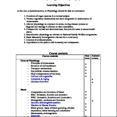
Physiology 6c6w
December 2019 81
Physiology 6c6w
December 2019 55
Membrane-bioreactor.ppt 37105z
December 2019 48
Membrane Processes 49335f
April 2022 0More Documents from "teklay" 66405a

Pediatric Vital Signs: Charts Of Normal Ranges l1k4o
December 2019 21
Chapter 1 Foundation Of Modern Nursing 2d632c
March 2021 0
Study Questions Endocrine System 1_dr P Kumar Biochem-genetics 3i5w3g
December 2019 27
Membrane Physiology 3w2a3j
March 2021 0
Tooth Nomenclature 6y5315
April 2022 0