Flap Surgery And Gingivectomy 1g665q
This document was ed by and they confirmed that they have the permission to share it. If you are author or own the copyright of this book, please report to us by using this report form. Report 2z6p3t
Overview 5o1f4z
& View Flap Surgery And Gingivectomy as PDF for free.
More details 6z3438
- Words: 7,489
- Pages: 127
|
V
Periodontal surgery comprises initial treatment, in which the original cause of periodontal disease is eliminated, and definitive surgery in which an environment conducive to long-term health and maintenance is achieved.
V
The decision concerning what type of periodontal surgery should be performed and how many sites should be included is usually made after the effect of initial cause-related measures has been evaluated.
!!" # $%!&$ &" !'!(!) V
›emoval of calculus and bacterial plaque will eliminate or markedly reduce the inflammatory cell infiltrate in the gingiva (edema, hyperemia, flabby tissue consistency), thereby making assessment of the ´trueµ gingival contours and pocket depths possible.
V
›eduction of gingival inflammation makes the soft tissues more fibrous and thus firmer, which facilitates surgical handling of the soft tissues. The propensity for bleeding is reduced, making inspection of the surgical field easier.
V
A better basis for a proper assessment of the prognosis has been established. The effectiveness of the patient·s home care, which is of decisive importance for the long-term prognosis, can be properly evaluated.
Ô
| V A flap is defined as a loosened section of tissue separated from the surrounding tissue except at its base. | V A periodontal flap is a section of gingiva and/or mucosa surgically separated from the underlying tissues to provide visibility of and access to the bone and root surface.
V
Traditionally, pocket elimination has been the main objective of periodontal therapy. The removal of the pocket by surgical means served two purposes:
(1) Elimination of pocket, which established an environment conducive to progression of periodontal disease. (2) the root surface was made accessible for scaling and for selfperformed tooth cleaning after healing. V
hile these objectives cannot be entirely discarded today, the necessity for pocket elimination in periodontal therapy has been challenged.
@ain objective of periodontal surgery is to contribute to the longterm preservation of the periodontium by facilitating plaque removal and infection control, and periodontal surgery can serve this purpose by: 1. Creating accessibility for proper professional scaling and root planing 2. Establishing a gingival morphology which facilitates the patient·s selfperformed infection control. V
thers -. ›egeneration of periodontal attachment lost due to destructive disease. 4. Elimination or reduction of pocket depth by resection of the pocket wall. 5. Expose the bone for osteoplasty or ressective osseous surgery 6. For root coverage procedures
á
?
# #
% &
!
"
$ $ % # $
)*! !!""%)*+# )*+""!! ' )'
' ' '
' ' ' '
( *
á + , - $ á $- $
% $ ) )
.
)
) " " " "
) # .$ # - # +
&' ) " ' * )
' ! ) $ )
Ô
!# ! V
The degree of access to the underlying bone and root surface necessary
V
The final position of the flap
V
idth of attached gingiva
V
Preservation of good blood supply to the flap is very important
',!$!" ith internal bevel incision
%*% *!"
ÿ, $!"
|)*+ V
f the flap is too thin, blood supply may be impaired and necrosis of the flap margin can occur, causing a slow and painful healing. f the flap is too thick, bulky tissue will result, leading to the formation of pseudopockets after healing.
deal thickness
Flap too thin
Necrosis of flap
i
iorizontal incisions are directed along the margin of the gingiva in a mesial or a distal direction. V V V
nternal bevel incision (primary incision) Sulcular incision (Secondary incision) nterdental incision (Third incision)
nternal bevel incision (›everse bevel): ectives: V t removes the pocket lining V t conserves the relatively uninvolved outer surface of the gingiva, which is apically positioned becomes attached gingiva. V Produces a sharp, thin flap margin for adaptation to bone tooth junction ndications
or internal evel incision
V Primary incision of flap surgery if there is a sufficient band of attached gingiva V Desire to correct bone morphology (osteoplasty, osseous resection) V Thick gingiva (such as palatal gingiva) V Deep periodontal pockets and bone defects V Desire to lengthen clinical crown
The internal bevel (first) incision can be made at varying locations and angles according to the different anatomic and pocket situations using blade no. 11 or 15.
An occlusal view of the different locations where the internal bevel incision can be made.
Second incision ² crevicular incision ndications
V Narrow band of attached gingiva V Thin gingiva and alveolar mucosa V Shallow periodontal pocket V Desire to lessen postoperative gingival recession for esthetic reasons in the maxillary anterior region. V As a second incision for flap surgery V Bone graft or GT›: desire to preserve as much periodontal tissue (interdental papilla) as possible to completely cover grafted bone and membrane by flaps. V
@ethod
V ncision given with beak shaped no. 12D blade the incision is carried around the entire teeth.
V V
V V
Third incision ² interdental incision This is made along the alveolar crest and alveolar septum from the buccal to the lingual side. This separates cervical secondary flap from the alveolar crest and interdental bone after reflecting the buccal and lingual flaps. @ethod The orban interdental knife is recommended
÷ if apical, lateral and coronal displacement of flap is not anticipated.
&!""*% -.%# !/0!&#"! , V V
V
V V
This consists of horizontal, internal bevel, non scalloped incision to remove the gingival papilla and denude the interdental space. Full-thickness flaps are elevated on both buccal and lingual. All granulation tissue is removed from all walls of the bony lesion and the bone and tooth are meticulously curetted. Some clinicians perforate the walls of the lesion with small round burs to allow migration of osteogenic elements from the adjacent marrow spaces. hen the flaps are replaced and sutured, the marginal bone is left denuded.The rationale is that it may take longer for the surface epithelium to migrate into the defect thus providing time for coronal regeneration to take place. Contraindicated when bone grafts used. iealing, if successful, occurs when bone is regenerated from the floor and walls of the lesion to fill the defect.
V
For full thickness flap blunt dissection is accomplished by periosteal elevation.
V
Sharp dissection is necessary to reflect a partial thickness flap, a surgical scalpel (no. 11 or no. 15) is used.
i ÷ ÷ ÷
þ þþ Neumann ie claimed the introduction of the mucoperiosteal flap in 1911. Neuman in 1912 and 1915 described a semilunar incision for access to the root surfaces and the alveolar crest. Later in Neuman·s text book in 1920 a mucoperiosteal periodontal flap was described the stressed that periodontal surgery results in horizontal alveolar and gingival atrophy.
1916 idman
Appears to be first to describe flap surgery for pocket elimination although Cieszynskin in a discussion in 1914 referred to periodontal flap for pocket reduction.idman introduced the idman flap in 1918.
1918 Cieszynski ie was credited with the introduction of reverse bevel incision. Zentler introduced the mucoperiosteal flap in the U.S.A. in 1918, with the idea that the procedure was to allow access for debridement and elimination of granulation tissue as well as osseous removal by chisels.
19-1 Kirkland
Apparently the first description of the flap procedure for the purpose of reattachment was given by Kirkland. iis method has been used as ´open subgingival curettage.µ
19-5 Kronfeld Flap surgery became popular after 19-5, when Kronfeld in his autopsy Orban study of which he stated that the bone adjacent to the periodontal pockets was neither necrotic nor infected but rather destroyed by an inflammatory process. Orban later ed this finding in his own studies. The main culprit of the disease now has shifted to the soft tissue. 1949 Schluger During 19-0 and 1940 gingivectomy was most popular for pocket elimination (Schluger in 1949). ie recommended gingivectomy followed by mucoperiosteal flap. Later a modification of Schluger·s approach:They were ¶pushback· and ¶pouch operation· with extensive exposure by alveolar process and mucobuccal fold extension following surgical remodeling of bone for pocket elimination. 1954 Nabers ie described the ´repositioning of the attached gingiva.µ 1957 Nabers
ie utilized the idman·s inverse bevel incision of which he called the ´repositioning incisionµ which includes the internal incision from the gingival margin to the alveolar crest.
Ô ! " #$%
**!"! "#-
& '( )
( ) V
An intracrevicular incision was made through the base of the gingival pockets and the entire gingiva (and part of the alveolar mucosa) was elevated in a mucoperiosteal flap.
V
Sectional releasing incisions were made to demarcate the area of surgery.
V
Following flap elevation, the inside of the flap was curetted to remove the pocket epithelium and the granulation tissue.
V
The root surfaces were subsequently carefully ´cleanedµ. Any irregularities of the alveolar bone were corrected to give the bone crest a horizontal outline.
V
The flaps were then trimmed to allow both an optimal adaptation to the teeth and a proper coverage of the alveolar bone on both the buccal/lingual (palatal) and the interproximal sites.
V
ith regard to pocket elimination, Neumann (1920) pointed out the importance of removing the soft tissue pockets, i.e. replacing the flap at the crest of the alveolar bone.
@) ) * )+ '1/'2 +#
Advantages V V V
V
There is not extensive sacrifice of non inflamed tissues Apical displacement of gingival margin is not done Since root surfaces were not markedly exposed the method could be esthetic, hence used in anterior regions of dentition. The potential for bone regeneration in intrabony defects.
, ) The choice of method is detected by: V
hether the osseous defects therapy is necessary or not.
V
The thickness of the gingiva and alveolar bone margin in the operative area.
V
f the periodontal pocket reaches or extends beyond the mucogingival junction and attached gingiva is extremely thin, the flap may be full thickness or partial thickness.
|%)*+
ndications V Pocket eradication V idening the zone of attached gingiva V crown lengthening procedures for cosmetic enhancement and restorative treatment Oontraindications V Compromised esthetic and anatomical preclusion
The split thickness A›P: ndications
V ncrease of the attached gingiva an area with narrow attached gingiva and sufficient oral vestibule depth with no extensive treatments for bone necessary V Avoid exposing areas where the alveolar bone is thin because of protruding tooth V where there is likelihood of osseous dehiscence or osseous fenestration V Elimination of the periodontal pocket that extends beyond the mucogingival junction with narrow attached gingiva. V Extension of clinical crown length for restorative/ prosthetic treatment (Crown lengthening surgery) V
Oontraindications V Thin gingiva V Lack of keratinized gingiva at gingival margin V Narrow oral vestibule V Extremely thin alveolar process V Extensive osseous surgery required V Deep intrabony defect requiring bone regeneration or restoration
*)3%
á
V
V
Advantages @inimum pocket depth post-operatively
V
f optimal soft tissue coverage of the alveolar bone is obtained, the post-surgical bone loss is minimal
V
The post-operative position of the gingival margin may be controlled and the entire muco-gingival complex may be maintained.
Disadvantages: V The sacrifice of periodontal tissues by bone resection and the subsequent exposure of root surfaces. V Technically demanding V Danger of penetrating flap during incision V Necrosis may result because of severe damage to blood vessels V Difficulty in manipulating the suture V Post operative swelling V Delayed healing because of secondary intention. V Treatment may be complicated if combined with osseous resection.
@) '( ) V
›amfjord and Nissle in 1974 coined the term modified idman flap though the procedure was employed by @orris in 1965 and was termed the unrepositioned mucoperiosteal flap
I )$ $ ' .
/ $ '
V
Technique
Advantages V V V V V
The possibility of obtaining a close adaptation of the soft tissues to the root surfaces. The minimum of trauma to which the alveolar bone and the soft connective tissues are exposed. Access for adequate instrumentation of root surfaces and immediate closure of the area @inimum trauma to which the alveolar bone and soft connective tissue are exposed Less exposure of the root surfaces which from an esthetic point of view is an advantage in the treatment of anterior segments of dentition.
@) '( ) V
V V
V V
Salaria S et al (2010) performed modified idman flap with the help of laser and called it Laser assisted @odified idman flap (LA@F). n the technique followed, the pocket lining was removed with the help of a diode laser. The fiberoptic tip of the laser was directed parallel to the root surface and was moved laterally and apically along the lateral pocket wall eventually reaching close to base of pocket. The laser settings were rearranged at 4 in continuous mode. 1.5 in continuous mode and applied in the sulcus for atleast 1 min to achieve laser bacterial reduction in the pocket as well as the connective tissue.
V V
V
V V
The laser was kept in continuous movement to prevent charring of connective tissue. Crevicular incision was given with a BP blade no. 15 directed towards the alveolar crest. Full thickness mucoperiosteal flap was raised buccally and lingually. The granulation tissue was removed from the defects by manual debridement and root surfaces were thoroughly planed. nner aspect of the reflected buccal and lingual flap was then subjected to laser application to remove any remaining epithelium. After complete debridement the surgical site was sutured. There was no pain or any other discomfort reported at 1 week post surgery.
)
-
- )$ '* $
$ '
V V V V V V V V V
The pockets are measured with periodontal probe and bleeding points are produced to mark bottom of the pocket. nternal bevel incision made and is carried to a point apical to alveolar crest. Thinning of flap is done with initial incision. Crevicular incision is made Flap is reflected with a periosteal elevator (blunt dissection) from internal bevel incision nterdental incision is made with interdental knife Triangular wedge is removed with curette Area is debrided and all tissue tags and granulation tissue is removed After scaling and root planing the flap edge should rest on the root bone junction A continuous sling suture is used to secure the facial and lingual flap to hold the flap edges at root bone junction. Area is covered with a periodontal pack.
) V
Because of anatomic characteristic of palate, palatal flap require different designs. The palatal tissue is masticatory mucosa and immobile. t is impossible to displace a palatal flap apically.
V
Close adaptation to the tooth surface and bone margin is difficult and post operative gingival morphology is unfavorable.
V
›eduction of periodontal pocket in a thick gingival wall in the palatal aspect is uncommon because of minimal gingival shrinkage achieved by initial therapy.
V
naccessibility of cleaning instruments may cause inadequate self care.
|!!- *!# #% !!" ""! *!# V
For debridement internal bevel incision planned so that flap adapts at root bone junction when sutured.
V
For osseous resection ² the incision should be planned to compensate for the lowered level of bone when flap is closed.
V
The initial incision varies with anatomic situation the internal bevel incision should be done in such a way that flap fits around tooth without exposing bone.
V
The initial incision is followed by crevicular and interdental incisions.
V
f tissue is thick horizontal gingivectomy incision made followed by internal bevel incision
V
Thick palatal should be thinned before reflection to adapt to the underlying osseous tissue and provide a thin knife edge margin.
V
The use of vertical incisions care must be taken to avoid the numerous vessels located in palate.
V
Ability to fix flap to optimal position by periosteal suture
) &- &, V V
Objective @aximum amount of gingival tissue and papilla are retained to cover the materials placed in the pocket.
! Technique V Step 1 using no. 12 blade incise the tissue at the bottom of the pocket and to the crest of bone, splitting the papilla below the point. V Step 2 reflect the flap maintaining it as thick as possible, not attempting to thin it as its done for ressective surgery. The maintenance of thick flap is necessary to prevent exposure of graft or membrane due to necrosis of flap. V
- )
V
ndications There must be adequate interdental space to allow the intact papilla to be reflected with facial and lingual flap.
V
Takei et al. (1985) proposed a surgical approach called papilla
preservation technique.
V
Cortellini et al. (1995, 1999) described modifications of the flap design to be used in combination with regenerative procedures.
V
For esthetic reasons, the papilla preservation technique is often utilized in the surgical treatment of anterior tooth regions.
V
) - V
@ "# $@% & & þ
V
developed in order to increase the space for regeneration, and in order to achieve and maintain primary closure of the flap in the interdental area
() - ) ierpaolo Oortellini, iovanpaolo ini rato, @aurizio
onetti,
)"*!|% -* !*)! ! !4* - $ !*#% !$5%**). !* ÿ
42 shows a deep pocket (PPD 15 mm) at the distal surface.The interdental space between 42 & 4- is very narrow as the result of rotation of 42. A high band of keratinized tissue is present at the buccal aspect of both teeth.
The buccal incision consists of the submarginal highly scalloped incisions at the two teeth neighboring the defect, the submarginal split-thickness incisions at the interdental papillae mesial and distal to the defect area, and one mesial vertical releasing incision.
A deep intrabony defect is evident after flap elevation and degranulation of the defect.
The membrane is positioned at the level of the bone crest and sutured at the periosteum left in place during buccal flap elevation. Note the space available above the membrane and below the area; this is adequate for soft tissue coverage of the membrane material.
Complete soft tissue coverage above the membrane has been achieved. At time of suture removal (14 days), the membrane was still covered.
At time of membrane removal (6 weeks), newly formed tissue completely fills the space available below the membrane. Note the good maturation of the regenerated tissue and the absence of clinical signs of inflammation.
Clinical aspect at 1 year. 42shows a shallow residual probing pocket (- mm) with minimal increase in the amount of gingival recession. A 2mm-high band of buccal keratinized tissue is still present at both teeth neighboring the defect area.
&$$% -**)3% ! 6! ÿ
# ! Ô%! ! !! Ô !! ! V V V
V V V
Techniques that induce the least amount of visual root exposure should be considered first. But nonelimination of the pocket may place the tooth in jeopardy. Final decision may have to be a compromise between health and esthetics, not attaining ideal results in either respect. Anterior teeth offer some advantages to a conservative approach. 1. They are all single rooted and easily accessible; 2. Patient's compliance and thoroughness in plaque control are easier to attain. Therefore scaling and root planing is the technique of choice for the anterior teeth.
V
V
V V
f surgery required for improved accessibility for root planing or regenerative surgery of osseous defects, papilla preservation flap can be used for both purposes and also offers a better postoperative result with less recession and reduced soft tissue crater formation interproximally. hen the teeth are too close interproximally, it may not be feasible, and sulcular incision flap offers good esthetic results and is the next choice. hen esthetics are not the primary consideration, @odified idman flap can be chosen. n some infrequent cases, bone contouring may be needed despite the resultant root exposure. The technique of choice is the apically displaced flap with bone contouring.
. V V
V
V V
Treatment for premolars and molars usually poses no esthetic problem but frequently involves difficult accessibility. Bone defects are more frequent and root morphologic features, particularly in relation to furcations, may offer unsurmountable problems for instrumentation in a close field. Therefore surgery is frequently indicated in this region. Accessibility can be obtained by either the undisplaced or apically displaced flap. @ost cases of moderate to severe periodontitis have developed osseous defects that require some degree of osseous remodeling or regenerative procedures.
V
hen osseous defects amenable to regeneration are present, the papilla preservation flap is the technique of choice because it better protects the interproximal areas where defects are frequently present.
V
Second and third choices are the sulcular flap and the modified idman flap, maintaining as much of the papilla as possible.
V
hen osseous defects with no possibility of reconstruction, such as interdental craters, are present, the technique of choice is the flap with osseous contouring.
Ô ! ! @" V V
V V
V V
Existing gingiva is preserved The marginal alveolar bone is exposed whereby the morphology of bony defects can be identified and the proper treatment rendered Furcation areas are exposed; the degree of involvement and the ´tooth²boneµ relationship can be identified The flap can be repositioned at its original level or shifted apically, thereby making it possible to adjust the gingival margin to the local conditions The flap procedure preserves the oral epithelium and often makes the use of surgical dressing superfluous The post-operative period is usually less unpleasant to the patient when compared to gingivectomy.
! @ Ô ! Ô # !Ô % / %! !"
@" Gingivectomy means excision of the gingiva.
! **+- !%# V
Ambroise Pare (1509-90) was the outstanding surgeon of the ›enaissance, and he developed many oral surgical procedures, including gingivectomy for hyperplastic gingival tissues.
V
The surgical approach as an alternative to sub - gingival scaling for pocket therapy was already recognized in the latter part of the nineteenth century, when ›obicsek (1884) pioneered the so-called gingivectomy procedure.
V
Pickerill $þ þ%was the first to use the term gingivectomy.
V
Gingivectomy was later defined by Grant et al. (1979) as being ´the excision of the soft tissue wall of a pathologic periodontal pocketµ. The surgical procedure, which aimed at ´pocket eliminationµ, was usually combined with recontouring of the diseased gingiva to restore physiologic form.
V
By removing the pocket wall, gingivectomy provides visibility and accessibility for complete calculus removal and thorough smoothing of the roots, creating a favorable environment for gingival healing and restoration of a physiologic gingival contour.
V
The gingivectomy technique was widely performed in the past.
V
mproved understanding of healing mechanisms and the development of more sophisticated flap methods have relegated the gingivectomy to a lesser role in the current repertoire of available techniques.
V
iowever, it remains an effective form of treatment when indicated.
V V
' ' Elimination of suprabony pockets, regardless of their depth, if the pocket wall is fibrous and firm.
V
Elimination of gingival enlargements.
V
Elimination of suprabony periodontal abscesses.
V
Pre-restorative caries exposure and crown lengthening in the treatment of subgingival carious lesions.
V V
'' ' 1. The need for bone surgery or examination of the bone shape and morphology
V
2. Situations in which the bottom of the pocket is apical to the mucogingival junction
V
-. Esthetic considerations, particularly in the anterior maxilla
!*+ 7'889:#. .5 7'1'8:#* #) --$*!& !*#% )"!!-&,
* / % ) 001'
* /2 0'
-$*!& !*#% !&#!#& #* #'1'&!#,
' ! ) ) ')3 ) /$ $ '* ) ) $ ) ) ) '
')* $ 4) 5 $ )$ '
* - 3 '
* $ $- '
) ' 6 $) ) '
* ) '
&-, V
Gingivoplasty is similar to gingivectomy, but its purpose is different.
V
Gingivoplasty is a reshaping of the gingiva to create physiologic gingival contours, with the sole purpose of recontouring the gingiva in the absence of pockets.
V
Gingival and periodontal disease often produce deformities in the gingiva that interfere with normal food excursion, collect plaque and food debris, and prolong and aggravate the disease process.
V
Gingival clefts and craters, shelf like interdental papillae caused by NUG, and gingival enlargement are examples of such deformities.
V
Gingivoplasty may be done with a periodontal knife, a scalpel, rotary coarse diamond stones, 6 or electrodes.
V
t consists of procedures that resemble those performed in festooning artificial dentures; namely, tapering the gingival margin creating a scalloped marginal outline thinning the attached gingiva creating vertical interdental grooves and shaping the interdental papillae to provide sluiceways for the age of food.
V V V V
&-(, , &, "# V The removal of gingival enlargements and gingivoplasty is performed with the needle electrode, supplemented by the small ovoid loop or the diamond-shaped electrodes for festooning. V
A blended cutting and coagulating (fully rectified) current is used. n all reshaping procedures, the electrode is activated and moved in a concise "shaving" motion.
V
For hemostasis, the ball electrode is used. iemorrhage must be controlled by direct pressure (via air, compress, or hemostat) first; then the surface is lightly touched with a coagulating current.
V
Electrosurgery is helpful for the control of isolated bleeding points. Bleeding areas located interproximally are reached with a thin, bar shaped electrode.
V
Electrosurgery permits an adequate contouring of the tissue.
V
V V
V
Controls hemorrhage.
V
V
Cannot be used in patients who have non compatible or poorly shielded cardiac pacemakers. The treatment causes an unpleasant odor. f the electrosurgery point touches the bone, irreparable damage can be done; The heat generated by injudicious use can cause tissue damage and loss of periodontal when the electrode is used close to bone. hen the electrode touches the root, areas of cementum burn are produced.
&&-(, V
The lasers most commonly used in dentistry are the CO2 and Nd:YAG, which have wavelengths of 10,600 nm and 1064 nm, respectively, both in the infrared range.
V
The CO2 laser beam has been used for the excision of gingival growths, although healing is delayed when compared with healing after conventional scalpel gingivectomy.
V
The use of laser beam for oral surgery requires precautionary measures to avoid reflecting the beam on instrument surfaces, which could result in injury to neighboring tissues and the eyes of the operator.
-$*!& &)!% - & V
Techniques to remove the gingiva using chemicals, such as 5% paraformaldehyde or potassium hydroxide, have been described in the past but are not currently used.
Disadvantages: 1. The depth of action cannot be controlled, and therefore healthy attached tissue underlying the pocket may be injured. 2. Gingival remodeling cannot be accomplished effectively. -. Epithelialization and reformation of the junctional epithelium and reestablishment of the alveolar crest fiber system are slower in chemically treated gingival wounds than in those produced by a scalpel . V
V
2
% @ %! !Ô # ! "
, ) ) & V
Primary wound healing (iealing by primary intention): occurs in incised wounds when minimal tissue loss has occurred and the wound edges are brought into by sutures. Because little, if any, granulation tissue is present, this type of wound heals rapidly, produces little structural or functional deficit, and involves minimal scarring.
V
Secondary wound healing (iealing by secondary intention): seen where the wound edges are widely separated either as a part of intentional surgical procedures or as a consequence of tissue loss or destruction. The defect becomes filled with granulation tissue, which is eventually remodeled to form a definite scar. Such repair requires restoration of epithelium and submucosa (or of dermis in skin wounds) by synthesis of new constituents. n patients with extensive tissue loss, secondary wound healing may result in significant functional or cosmetic problems with excessive scar formation.
V
V
Third intention wound healing (Delayed primary closure): in healing by third intention, the wound is temporarily left open, usually because of contamination.The wound is then closed after 4 to 7 days, with wound approximation being accomplished by either grafting or flap rotation.
#& )0& &-(, nitial iealing (0-5 iours)
V V
V
5 iours - 1 Day Epithelium: A distinct movement of epithelial cells that originates at the wound margin begins to cross the surface of the wound.This migration is from the spinous layer and occurs by cells wedging themselves between the surface coagulum and the underlying viable connective tissue of the wound. Epithelial proliferation has also begun at the wound edge, as seen by the increased presence of mitotic figures. Connective tissue: Proliferation begins beneath and within the polyband.The clot and polyband serve a protective function until the epithelium forms a continuous sheath over the wound.The clot and P@Ns disappear later when the epithelial sheath is formed.
V V
2-- Days Epithelial cells are covering the wound at a rate of 0.5 mm/day.The smoother the surface of the connective tissue after the surgical procedure, the faster the rate of epithelialization. Both basal cells of the wound margin and cells that have migrated onto the wound surface are proliferating.There is a decrease in inflammatory cells (P@Ns) below the migrating epithelial cells.
V
0-4 days
D
10-28 Days
( ) 1 &, #& f a long bevel is made (exposing a greater wound surface), then the healing will take longer because the epithelial cells have further to migrate. The more accurate the bevel is to 45º, the less height in the regenerated gingival margin (especially when a thick gingival ledge is seen).
-" * !% - & V
Some investigators report no significant differences in gingival healing after resection by electrosurgery and resection with periodontal knives; other researchers find delayed healing, greater reduction in gingival height, and more bone injury after electrosurgery.
V
There appears to be little difference in the results obtained after shallow gingival resection with electrosurgery and that with periodontal knives.
V
iowever, when used
or deep resections close to one,
electrosurgery can produce gingival recession, one necrosis and
sequestration, loss o one height,
urcation exposure, and tooth
moility, which do not occur with the use o periodontal knives.
-" --$*!& V
V
V V
The acute inflammatory reaction is delayed and minimal, and few myofibroblasts are present in the base of the wound during healing. These cells are the effectors of wound contraction and thus there is little contraction following removal of oral mucosa with laser. There is minimal scarring, resulting in little duration or restriction in movement of the soft tissues. There is less bleeding with laser as compared to the scalpel cut, since the blood vessels of .5mm or less in diameter are sealed spontaneously, thus aided to the surgeon visibility and precision during the surgery.
V
Disadvantage: @ild postoperative discomfort usually delayed up to one or two weeks. Epithelial regeneration is delayed, and the wounds take a longer time to reepithelialize than following conventional surgery. Another disadvantage is the high cost of equipment.
V
This delayed of wound healing is thought to be the result of the lack of wound contraction and thus failure of the area to reduce in size. The procedure usually will take longer time especially in gingivoplasty where abundant of tissue removal is required.
à Ô Ô
#& ) ) &, V V
V
V
nitial iealing (1 - - Days) Space between flap and tooth or bone is thinner, epithelial cells migrate over the border of the flap. rreversible bone resorption is greatest in areas where alveolar bone is thin and is comprised of two plates of compact bone fused together (e.g. - over prominent roots and the labial areas of mandibular incisors). Less irreversible destruction of bone occurs where cancellous or ing bone exists between the outer cortical plate and alveolar bone proper because the marrow spaces and vascular sites act as a reservoir for healing.
09#&
90' #&
' 0ÿ'#&
ÿ'#& 21 Days
One month after surgery: A fully epithelized gingival crevice with well defined epithelial attachment is present. There is a beginning junctional arrangement of supracrestal fibers.
& V
V
V
V
V
The epithelial adhesion occlusal to the base of the original pocket is seen at times, but more frequently the collagen adhesion immediately apical to the newly adhering junctional epithelium but occlusal to the marginal alveolar crest. n the infrabony portion of the periodontal lesions, simultaneous and/or sequential deposition of repair cementum, functionally oriented ligament fibers and narrowing of the vertical defect by osteogenesis. t is possible that supracrestal healing following flap surgery is a connective tissue adherence over a limited space immediately apical to the junctional epithelial adherence. Another pattern of healing in connective tissue is the splicing of fiber ends from the tooth surface with new fibers from the healing flap wound edge. This occurs when the root surface is not completely devoid of periodontal tissue (whether due to pathological lesion or mechanical/chemical debridement). This type of healing can be classified as repair rather than regeneration.
V
V
V
V
V
iistologic observation also has indicated that there may be a newly cementum like material to be deposited against the tooth coronal to the crest.This newly deposited cementum is cellular, not unlike bone, and be best described as repair cementum (Dragoo and Sullivan 197-, iawley and @iller 1975). Listgarten (1972) stated that this cementum is devoid of well defined fiber bundle. Again, since the new regenerative tissue is not exact the same as the original, the healing process can best be described as repair rather than regeneration. Theodore i. Dedolph and ienry B. Clark in 1958 revealed that at - weeks the epithelial attachment was complete, the attachement of periodontal membrane fibers and other connective tissue elements was restored, and the inflammatory response was mild or absent. n the study in monkey by Caffesse and ›amfjord and Nasjletti in 1968, healing following reverse bevel periodontal flap surgery has been characterized by first, second, or third intention, depending on the state of flap adaptation to teeth. Finally, the transient lowering of the attachment level and bone resorption at the alveolar crest - to 4 weeks following flap surgery tend to heal back to the presurgical level within 10 weeks after the surgery.
, )
Ô
Ô
() '( )
Ô
Ô
2 ) ) &, V
V
V V
V
Full thickness flap which denudes the bone result in superficial bone necrosis at 1 to - days, osteoclastic resorption follows and reach a peak at 4 ² 5 days, declining thereafter. During this dissective procedure, the periosteum is usually separated from the alveolar bone, initiating a striking remodeling activity known as " ( $%, which hastens the healing process (Yaffe A et al 1994). The phenomenon is a transient burst of localized remodeling activity following surgical wounding of cortical bone. This results in a loss of bone about 1mm, bone loss in greater if bone is thin.Various reports state that periodontal flap surgery results in varying amounts of alveolar crest loss (ood DL et al 1972) and interdental bone loss (Bragger U et al 1988). This bone loss may range from a minimum of 0.11mm to a maximum of -.1mm with an average of 0.8mm (ilderman et al 1970).
V
Osteoplasty (thinning of the buccal bone) using diamond burs, included as part of the surgical technique, results in areas of bone necrosis with reduction in bone height, which is later remodeled by new bone formation.
V
Therefore the final shape of the crest is determined more by osseous remodeling than by surgical reshaping.
V
Bone repair reaches its peak at - to 4 weeks. Loss of bone occurs in the initial healing stages both in radicular bone and in interdental bone areas.
V
iowever, in interdental areas, which have cancellous bone, the subsequent repair stage results in total restitution without any loss of bone; whereas in radicular bone, particularly if thin and uned by cancellous bone, bone repair results in loss of marginal bone.
! -!!" "%)*+ # )*+" V
Study in 1972 by Dale L. ood, Phillip ioag, O. alter Donnenfeld, and Leon ›osenfeld reveals of the loss of crestal radicular bone after both the full thickness and the partial thickness flap. The mean bone loss for the full thickness flap was .62 mm and after partial thickness flaps was .98mm. So, bone loss was greater with the use of partial thickness flap.
V
Fickl S et al 2011: Partial-thickness flaps resulted in less bone loss than full-thickness flaps, but are subject to some variability. Use of partial-thickness flaps does not prevent from all bone loss.
) 2 2 )0& ) &, V
n earlier studies, it has been proved that bisphosphonate alendronate (ALN) is effective in reducing alveolar bone loss following periodontal surgery in rats. Topical application of sponge soaked in 10 Ul of ALN solution at the time of surgery demonstrated marked reduction of bone resorption while maintaining the alveolar crest height (Yaffe A et al 1997).
V
Aminobisphosphonate given intravenously significantly reduced alveolar bone resorption after mucoperiosteal flap surgery (Yaffe A et al 1995).
V
Because of the high affinity of bisphosphonates for iA bone mineral, these drugs are targeted to areas of bone turnover and are specifically concentrated at the site of osteoclastic bone resorption.
V
Therefore, the most likely route by which bisphosphonates could inhibit bone resorption is by a direct effect on resorbing osteoclasts, though the fine mechanism/mechanisms by which bisphosphonates act on bone are not very clear.
V
There may be an indirect effect on secretion of osteoclast activating factor by osteoblasts.
V
The direct effect on osteoclasts includes cytotoxic or metabolic injury of mature osteoclasts, inhibition of osteoclast attachment to bone, inhibition of osteoclast differentiation or recruitment, and interference with osteoclast structural features necessary for bone resorption.
!*% *) V
Bisphosphonates bind to the bone mineral & are released due to highly acidic local environment.
V
Taken up by osteoclasts. nside the osteoclasts, they inhibit the enzyme(s) of the mevalonate pathway responsible for the production of cholesterol and isoprenoid lipids which are important substrates for the post-translational lipid modification (prenylation) of small GTPases.
V
Prenylation is required for the correct functioning of these proteins. nhibition of the mevalonate pathway and thus interference with generation of GGPP results in loss of prenylation of these GTP binding proteins. This results in disruption of actin cytoskeleton and loss of ruffled border formation, both of which are essential for the bone resorbing activity of osteoclasts, resulting in osteoclast inactivation.
V
V
!.)!)!."*!#)! - $ .&))% - &0#%*##)** !$) "! *!"- ",%# V
V
V
V
n 1995, ›eddy et al. also evaluated ALN for inhibition of alveolar bone loss in naturally occurring periodontitis. The results demonstrated that though there were no differences in signs of inflammation or pocketing, there was still an increase in bone mineral density. @eraw et al. 1999 conducted a study to evaluate the effect of local delivery of ALN on bone regeneration within peri-implant defects. The results indicated that ALN increases the amount of peripheral periimplant bone. Kaynak et al. 2000 in their study concluded that local application of ALN could be used as an adjunct in reducing bone resorption following surgery. Jyoti Gupta et al (2011) evaluated the relative efficacy of the alloplast used alone and in conjunction with an osteoclast inhibitor (topically delivered bisphosphonate) in the treatment of human periodontal infrabony defects. The results of the study show evidence of aminobispho-sphonates potentiating osseous regeneration by virtue of their osteoclast inhibitory activity, following mucoperiosteal flap surgery.
V V
Cheng A et al (2009) The possibility that bisphosphonates may be a useful treatment in periodontics was contemplated in the 1990s and early 2000s.
V
t was considered that bisphosphonate use may prevent bone loss around teeth and implants and possibly stimulate new bone formation.
V
These studies all ceased once osteonecrosis of the jaws ONJ was described in the literature.
%
V
Periodontal surgery comprises initial treatment, in which the original cause of periodontal disease is eliminated, and definitive surgery in which an environment conducive to long-term health and maintenance is achieved.
V
The decision concerning what type of periodontal surgery should be performed and how many sites should be included is usually made after the effect of initial cause-related measures has been evaluated.
!!" # $%!&$ &" !'!(!) V
›emoval of calculus and bacterial plaque will eliminate or markedly reduce the inflammatory cell infiltrate in the gingiva (edema, hyperemia, flabby tissue consistency), thereby making assessment of the ´trueµ gingival contours and pocket depths possible.
V
›eduction of gingival inflammation makes the soft tissues more fibrous and thus firmer, which facilitates surgical handling of the soft tissues. The propensity for bleeding is reduced, making inspection of the surgical field easier.
V
A better basis for a proper assessment of the prognosis has been established. The effectiveness of the patient·s home care, which is of decisive importance for the long-term prognosis, can be properly evaluated.
Ô
| V A flap is defined as a loosened section of tissue separated from the surrounding tissue except at its base. | V A periodontal flap is a section of gingiva and/or mucosa surgically separated from the underlying tissues to provide visibility of and access to the bone and root surface.
V
Traditionally, pocket elimination has been the main objective of periodontal therapy. The removal of the pocket by surgical means served two purposes:
(1) Elimination of pocket, which established an environment conducive to progression of periodontal disease. (2) the root surface was made accessible for scaling and for selfperformed tooth cleaning after healing. V
hile these objectives cannot be entirely discarded today, the necessity for pocket elimination in periodontal therapy has been challenged.
@ain objective of periodontal surgery is to contribute to the longterm preservation of the periodontium by facilitating plaque removal and infection control, and periodontal surgery can serve this purpose by: 1. Creating accessibility for proper professional scaling and root planing 2. Establishing a gingival morphology which facilitates the patient·s selfperformed infection control. V
thers -. ›egeneration of periodontal attachment lost due to destructive disease. 4. Elimination or reduction of pocket depth by resection of the pocket wall. 5. Expose the bone for osteoplasty or ressective osseous surgery 6. For root coverage procedures
á
?
# #
% &
!
"
$ $ % # $
)*! !!""%)*+# )*+""!! ' )'
' ' '
' ' ' '
( *
á + , - $ á $- $
% $ ) )
.
)
) " " " "
) # .$ # - # +
&' ) " ' * )
' ! ) $ )
Ô
!# ! V
The degree of access to the underlying bone and root surface necessary
V
The final position of the flap
V
idth of attached gingiva
V
Preservation of good blood supply to the flap is very important
',!$!" ith internal bevel incision
%*% *!"
ÿ, $!"
|)*+ V
f the flap is too thin, blood supply may be impaired and necrosis of the flap margin can occur, causing a slow and painful healing. f the flap is too thick, bulky tissue will result, leading to the formation of pseudopockets after healing.
deal thickness
Flap too thin
Necrosis of flap
i
iorizontal incisions are directed along the margin of the gingiva in a mesial or a distal direction. V V V
nternal bevel incision (primary incision) Sulcular incision (Secondary incision) nterdental incision (Third incision)
nternal bevel incision (›everse bevel): ectives: V t removes the pocket lining V t conserves the relatively uninvolved outer surface of the gingiva, which is apically positioned becomes attached gingiva. V Produces a sharp, thin flap margin for adaptation to bone tooth junction ndications
or internal evel incision
V Primary incision of flap surgery if there is a sufficient band of attached gingiva V Desire to correct bone morphology (osteoplasty, osseous resection) V Thick gingiva (such as palatal gingiva) V Deep periodontal pockets and bone defects V Desire to lengthen clinical crown
The internal bevel (first) incision can be made at varying locations and angles according to the different anatomic and pocket situations using blade no. 11 or 15.
An occlusal view of the different locations where the internal bevel incision can be made.
Second incision ² crevicular incision ndications
V Narrow band of attached gingiva V Thin gingiva and alveolar mucosa V Shallow periodontal pocket V Desire to lessen postoperative gingival recession for esthetic reasons in the maxillary anterior region. V As a second incision for flap surgery V Bone graft or GT›: desire to preserve as much periodontal tissue (interdental papilla) as possible to completely cover grafted bone and membrane by flaps. V
@ethod
V ncision given with beak shaped no. 12D blade the incision is carried around the entire teeth.
V V
V V
Third incision ² interdental incision This is made along the alveolar crest and alveolar septum from the buccal to the lingual side. This separates cervical secondary flap from the alveolar crest and interdental bone after reflecting the buccal and lingual flaps. @ethod The orban interdental knife is recommended
÷ if apical, lateral and coronal displacement of flap is not anticipated.
&!""*% -.%# !/0!&#"! , V V
V
V V
This consists of horizontal, internal bevel, non scalloped incision to remove the gingival papilla and denude the interdental space. Full-thickness flaps are elevated on both buccal and lingual. All granulation tissue is removed from all walls of the bony lesion and the bone and tooth are meticulously curetted. Some clinicians perforate the walls of the lesion with small round burs to allow migration of osteogenic elements from the adjacent marrow spaces. hen the flaps are replaced and sutured, the marginal bone is left denuded.The rationale is that it may take longer for the surface epithelium to migrate into the defect thus providing time for coronal regeneration to take place. Contraindicated when bone grafts used. iealing, if successful, occurs when bone is regenerated from the floor and walls of the lesion to fill the defect.
V
For full thickness flap blunt dissection is accomplished by periosteal elevation.
V
Sharp dissection is necessary to reflect a partial thickness flap, a surgical scalpel (no. 11 or no. 15) is used.
i ÷ ÷ ÷
þ þþ Neumann ie claimed the introduction of the mucoperiosteal flap in 1911. Neuman in 1912 and 1915 described a semilunar incision for access to the root surfaces and the alveolar crest. Later in Neuman·s text book in 1920 a mucoperiosteal periodontal flap was described the stressed that periodontal surgery results in horizontal alveolar and gingival atrophy.
1916 idman
Appears to be first to describe flap surgery for pocket elimination although Cieszynskin in a discussion in 1914 referred to periodontal flap for pocket reduction.idman introduced the idman flap in 1918.
1918 Cieszynski ie was credited with the introduction of reverse bevel incision. Zentler introduced the mucoperiosteal flap in the U.S.A. in 1918, with the idea that the procedure was to allow access for debridement and elimination of granulation tissue as well as osseous removal by chisels.
19-1 Kirkland
Apparently the first description of the flap procedure for the purpose of reattachment was given by Kirkland. iis method has been used as ´open subgingival curettage.µ
19-5 Kronfeld Flap surgery became popular after 19-5, when Kronfeld in his autopsy Orban study of which he stated that the bone adjacent to the periodontal pockets was neither necrotic nor infected but rather destroyed by an inflammatory process. Orban later ed this finding in his own studies. The main culprit of the disease now has shifted to the soft tissue. 1949 Schluger During 19-0 and 1940 gingivectomy was most popular for pocket elimination (Schluger in 1949). ie recommended gingivectomy followed by mucoperiosteal flap. Later a modification of Schluger·s approach:They were ¶pushback· and ¶pouch operation· with extensive exposure by alveolar process and mucobuccal fold extension following surgical remodeling of bone for pocket elimination. 1954 Nabers ie described the ´repositioning of the attached gingiva.µ 1957 Nabers
ie utilized the idman·s inverse bevel incision of which he called the ´repositioning incisionµ which includes the internal incision from the gingival margin to the alveolar crest.
Ô ! " #$%
**!"! "#-
& '( )
( ) V
An intracrevicular incision was made through the base of the gingival pockets and the entire gingiva (and part of the alveolar mucosa) was elevated in a mucoperiosteal flap.
V
Sectional releasing incisions were made to demarcate the area of surgery.
V
Following flap elevation, the inside of the flap was curetted to remove the pocket epithelium and the granulation tissue.
V
The root surfaces were subsequently carefully ´cleanedµ. Any irregularities of the alveolar bone were corrected to give the bone crest a horizontal outline.
V
The flaps were then trimmed to allow both an optimal adaptation to the teeth and a proper coverage of the alveolar bone on both the buccal/lingual (palatal) and the interproximal sites.
V
ith regard to pocket elimination, Neumann (1920) pointed out the importance of removing the soft tissue pockets, i.e. replacing the flap at the crest of the alveolar bone.
@) ) * )+ '1/'2 +#
Advantages V V V
V
There is not extensive sacrifice of non inflamed tissues Apical displacement of gingival margin is not done Since root surfaces were not markedly exposed the method could be esthetic, hence used in anterior regions of dentition. The potential for bone regeneration in intrabony defects.
, ) The choice of method is detected by: V
hether the osseous defects therapy is necessary or not.
V
The thickness of the gingiva and alveolar bone margin in the operative area.
V
f the periodontal pocket reaches or extends beyond the mucogingival junction and attached gingiva is extremely thin, the flap may be full thickness or partial thickness.
|%)*+
ndications V Pocket eradication V idening the zone of attached gingiva V crown lengthening procedures for cosmetic enhancement and restorative treatment Oontraindications V Compromised esthetic and anatomical preclusion
The split thickness A›P: ndications
V ncrease of the attached gingiva an area with narrow attached gingiva and sufficient oral vestibule depth with no extensive treatments for bone necessary V Avoid exposing areas where the alveolar bone is thin because of protruding tooth V where there is likelihood of osseous dehiscence or osseous fenestration V Elimination of the periodontal pocket that extends beyond the mucogingival junction with narrow attached gingiva. V Extension of clinical crown length for restorative/ prosthetic treatment (Crown lengthening surgery) V
Oontraindications V Thin gingiva V Lack of keratinized gingiva at gingival margin V Narrow oral vestibule V Extremely thin alveolar process V Extensive osseous surgery required V Deep intrabony defect requiring bone regeneration or restoration
*)3%
á
V
V
Advantages @inimum pocket depth post-operatively
V
f optimal soft tissue coverage of the alveolar bone is obtained, the post-surgical bone loss is minimal
V
The post-operative position of the gingival margin may be controlled and the entire muco-gingival complex may be maintained.
Disadvantages: V The sacrifice of periodontal tissues by bone resection and the subsequent exposure of root surfaces. V Technically demanding V Danger of penetrating flap during incision V Necrosis may result because of severe damage to blood vessels V Difficulty in manipulating the suture V Post operative swelling V Delayed healing because of secondary intention. V Treatment may be complicated if combined with osseous resection.
@) '( ) V
›amfjord and Nissle in 1974 coined the term modified idman flap though the procedure was employed by @orris in 1965 and was termed the unrepositioned mucoperiosteal flap
I )$ $ ' .
/ $ '
V
Technique
Advantages V V V V V
The possibility of obtaining a close adaptation of the soft tissues to the root surfaces. The minimum of trauma to which the alveolar bone and the soft connective tissues are exposed. Access for adequate instrumentation of root surfaces and immediate closure of the area @inimum trauma to which the alveolar bone and soft connective tissue are exposed Less exposure of the root surfaces which from an esthetic point of view is an advantage in the treatment of anterior segments of dentition.
@) '( ) V
V V
V V
Salaria S et al (2010) performed modified idman flap with the help of laser and called it Laser assisted @odified idman flap (LA@F). n the technique followed, the pocket lining was removed with the help of a diode laser. The fiberoptic tip of the laser was directed parallel to the root surface and was moved laterally and apically along the lateral pocket wall eventually reaching close to base of pocket. The laser settings were rearranged at 4 in continuous mode. 1.5 in continuous mode and applied in the sulcus for atleast 1 min to achieve laser bacterial reduction in the pocket as well as the connective tissue.
V V
V
V V
The laser was kept in continuous movement to prevent charring of connective tissue. Crevicular incision was given with a BP blade no. 15 directed towards the alveolar crest. Full thickness mucoperiosteal flap was raised buccally and lingually. The granulation tissue was removed from the defects by manual debridement and root surfaces were thoroughly planed. nner aspect of the reflected buccal and lingual flap was then subjected to laser application to remove any remaining epithelium. After complete debridement the surgical site was sutured. There was no pain or any other discomfort reported at 1 week post surgery.
)
-
- )$ '* $
$ '
V V V V V V V V V
The pockets are measured with periodontal probe and bleeding points are produced to mark bottom of the pocket. nternal bevel incision made and is carried to a point apical to alveolar crest. Thinning of flap is done with initial incision. Crevicular incision is made Flap is reflected with a periosteal elevator (blunt dissection) from internal bevel incision nterdental incision is made with interdental knife Triangular wedge is removed with curette Area is debrided and all tissue tags and granulation tissue is removed After scaling and root planing the flap edge should rest on the root bone junction A continuous sling suture is used to secure the facial and lingual flap to hold the flap edges at root bone junction. Area is covered with a periodontal pack.
) V
Because of anatomic characteristic of palate, palatal flap require different designs. The palatal tissue is masticatory mucosa and immobile. t is impossible to displace a palatal flap apically.
V
Close adaptation to the tooth surface and bone margin is difficult and post operative gingival morphology is unfavorable.
V
›eduction of periodontal pocket in a thick gingival wall in the palatal aspect is uncommon because of minimal gingival shrinkage achieved by initial therapy.
V
naccessibility of cleaning instruments may cause inadequate self care.
|!!- *!# #% !!" ""! *!# V
For debridement internal bevel incision planned so that flap adapts at root bone junction when sutured.
V
For osseous resection ² the incision should be planned to compensate for the lowered level of bone when flap is closed.
V
The initial incision varies with anatomic situation the internal bevel incision should be done in such a way that flap fits around tooth without exposing bone.
V
The initial incision is followed by crevicular and interdental incisions.
V
f tissue is thick horizontal gingivectomy incision made followed by internal bevel incision
V
Thick palatal should be thinned before reflection to adapt to the underlying osseous tissue and provide a thin knife edge margin.
V
The use of vertical incisions care must be taken to avoid the numerous vessels located in palate.
V
Ability to fix flap to optimal position by periosteal suture
) &- &, V V
Objective @aximum amount of gingival tissue and papilla are retained to cover the materials placed in the pocket.
! Technique V Step 1 using no. 12 blade incise the tissue at the bottom of the pocket and to the crest of bone, splitting the papilla below the point. V Step 2 reflect the flap maintaining it as thick as possible, not attempting to thin it as its done for ressective surgery. The maintenance of thick flap is necessary to prevent exposure of graft or membrane due to necrosis of flap. V
- )
V
ndications There must be adequate interdental space to allow the intact papilla to be reflected with facial and lingual flap.
V
Takei et al. (1985) proposed a surgical approach called papilla
preservation technique.
V
Cortellini et al. (1995, 1999) described modifications of the flap design to be used in combination with regenerative procedures.
V
For esthetic reasons, the papilla preservation technique is often utilized in the surgical treatment of anterior tooth regions.
V
) - V
@ "# $@% & & þ
V
developed in order to increase the space for regeneration, and in order to achieve and maintain primary closure of the flap in the interdental area
() - ) ierpaolo Oortellini, iovanpaolo ini rato, @aurizio
onetti,
)"*!|% -* !*)! ! !4* - $ !*#% !$5%**). !* ÿ
42 shows a deep pocket (PPD 15 mm) at the distal surface.The interdental space between 42 & 4- is very narrow as the result of rotation of 42. A high band of keratinized tissue is present at the buccal aspect of both teeth.
The buccal incision consists of the submarginal highly scalloped incisions at the two teeth neighboring the defect, the submarginal split-thickness incisions at the interdental papillae mesial and distal to the defect area, and one mesial vertical releasing incision.
A deep intrabony defect is evident after flap elevation and degranulation of the defect.
The membrane is positioned at the level of the bone crest and sutured at the periosteum left in place during buccal flap elevation. Note the space available above the membrane and below the area; this is adequate for soft tissue coverage of the membrane material.
Complete soft tissue coverage above the membrane has been achieved. At time of suture removal (14 days), the membrane was still covered.
At time of membrane removal (6 weeks), newly formed tissue completely fills the space available below the membrane. Note the good maturation of the regenerated tissue and the absence of clinical signs of inflammation.
Clinical aspect at 1 year. 42shows a shallow residual probing pocket (- mm) with minimal increase in the amount of gingival recession. A 2mm-high band of buccal keratinized tissue is still present at both teeth neighboring the defect area.
&$$% -**)3% ! 6! ÿ
# ! Ô%! ! !! Ô !! ! V V V
V V V
Techniques that induce the least amount of visual root exposure should be considered first. But nonelimination of the pocket may place the tooth in jeopardy. Final decision may have to be a compromise between health and esthetics, not attaining ideal results in either respect. Anterior teeth offer some advantages to a conservative approach. 1. They are all single rooted and easily accessible; 2. Patient's compliance and thoroughness in plaque control are easier to attain. Therefore scaling and root planing is the technique of choice for the anterior teeth.
V
V
V V
f surgery required for improved accessibility for root planing or regenerative surgery of osseous defects, papilla preservation flap can be used for both purposes and also offers a better postoperative result with less recession and reduced soft tissue crater formation interproximally. hen the teeth are too close interproximally, it may not be feasible, and sulcular incision flap offers good esthetic results and is the next choice. hen esthetics are not the primary consideration, @odified idman flap can be chosen. n some infrequent cases, bone contouring may be needed despite the resultant root exposure. The technique of choice is the apically displaced flap with bone contouring.
. V V
V
V V
Treatment for premolars and molars usually poses no esthetic problem but frequently involves difficult accessibility. Bone defects are more frequent and root morphologic features, particularly in relation to furcations, may offer unsurmountable problems for instrumentation in a close field. Therefore surgery is frequently indicated in this region. Accessibility can be obtained by either the undisplaced or apically displaced flap. @ost cases of moderate to severe periodontitis have developed osseous defects that require some degree of osseous remodeling or regenerative procedures.
V
hen osseous defects amenable to regeneration are present, the papilla preservation flap is the technique of choice because it better protects the interproximal areas where defects are frequently present.
V
Second and third choices are the sulcular flap and the modified idman flap, maintaining as much of the papilla as possible.
V
hen osseous defects with no possibility of reconstruction, such as interdental craters, are present, the technique of choice is the flap with osseous contouring.
Ô ! ! @" V V
V V
V V
Existing gingiva is preserved The marginal alveolar bone is exposed whereby the morphology of bony defects can be identified and the proper treatment rendered Furcation areas are exposed; the degree of involvement and the ´tooth²boneµ relationship can be identified The flap can be repositioned at its original level or shifted apically, thereby making it possible to adjust the gingival margin to the local conditions The flap procedure preserves the oral epithelium and often makes the use of surgical dressing superfluous The post-operative period is usually less unpleasant to the patient when compared to gingivectomy.
! @ Ô ! Ô # !Ô % / %! !"
@" Gingivectomy means excision of the gingiva.
! **+- !%# V
Ambroise Pare (1509-90) was the outstanding surgeon of the ›enaissance, and he developed many oral surgical procedures, including gingivectomy for hyperplastic gingival tissues.
V
The surgical approach as an alternative to sub - gingival scaling for pocket therapy was already recognized in the latter part of the nineteenth century, when ›obicsek (1884) pioneered the so-called gingivectomy procedure.
V
Pickerill $þ þ%was the first to use the term gingivectomy.
V
Gingivectomy was later defined by Grant et al. (1979) as being ´the excision of the soft tissue wall of a pathologic periodontal pocketµ. The surgical procedure, which aimed at ´pocket eliminationµ, was usually combined with recontouring of the diseased gingiva to restore physiologic form.
V
By removing the pocket wall, gingivectomy provides visibility and accessibility for complete calculus removal and thorough smoothing of the roots, creating a favorable environment for gingival healing and restoration of a physiologic gingival contour.
V
The gingivectomy technique was widely performed in the past.
V
mproved understanding of healing mechanisms and the development of more sophisticated flap methods have relegated the gingivectomy to a lesser role in the current repertoire of available techniques.
V
iowever, it remains an effective form of treatment when indicated.
V V
' ' Elimination of suprabony pockets, regardless of their depth, if the pocket wall is fibrous and firm.
V
Elimination of gingival enlargements.
V
Elimination of suprabony periodontal abscesses.
V
Pre-restorative caries exposure and crown lengthening in the treatment of subgingival carious lesions.
V V
'' ' 1. The need for bone surgery or examination of the bone shape and morphology
V
2. Situations in which the bottom of the pocket is apical to the mucogingival junction
V
-. Esthetic considerations, particularly in the anterior maxilla
!*+ 7'889:#. .5 7'1'8:#* #) --$*!& !*#% )"!!-&,
* / % ) 001'
* /2 0'
-$*!& !*#% !&#!#& #* #'1'&!#,
' ! ) ) ')3 ) /$ $ '* ) ) $ ) ) ) '
')* $ 4) 5 $ )$ '
* - 3 '
* $ $- '
) ' 6 $) ) '
* ) '
&-, V
Gingivoplasty is similar to gingivectomy, but its purpose is different.
V
Gingivoplasty is a reshaping of the gingiva to create physiologic gingival contours, with the sole purpose of recontouring the gingiva in the absence of pockets.
V
Gingival and periodontal disease often produce deformities in the gingiva that interfere with normal food excursion, collect plaque and food debris, and prolong and aggravate the disease process.
V
Gingival clefts and craters, shelf like interdental papillae caused by NUG, and gingival enlargement are examples of such deformities.
V
Gingivoplasty may be done with a periodontal knife, a scalpel, rotary coarse diamond stones, 6 or electrodes.
V
t consists of procedures that resemble those performed in festooning artificial dentures; namely, tapering the gingival margin creating a scalloped marginal outline thinning the attached gingiva creating vertical interdental grooves and shaping the interdental papillae to provide sluiceways for the age of food.
V V V V
&-(, , &, "# V The removal of gingival enlargements and gingivoplasty is performed with the needle electrode, supplemented by the small ovoid loop or the diamond-shaped electrodes for festooning. V
A blended cutting and coagulating (fully rectified) current is used. n all reshaping procedures, the electrode is activated and moved in a concise "shaving" motion.
V
For hemostasis, the ball electrode is used. iemorrhage must be controlled by direct pressure (via air, compress, or hemostat) first; then the surface is lightly touched with a coagulating current.
V
Electrosurgery is helpful for the control of isolated bleeding points. Bleeding areas located interproximally are reached with a thin, bar shaped electrode.
V
Electrosurgery permits an adequate contouring of the tissue.
V
V V
V
Controls hemorrhage.
V
V
Cannot be used in patients who have non compatible or poorly shielded cardiac pacemakers. The treatment causes an unpleasant odor. f the electrosurgery point touches the bone, irreparable damage can be done; The heat generated by injudicious use can cause tissue damage and loss of periodontal when the electrode is used close to bone. hen the electrode touches the root, areas of cementum burn are produced.
&&-(, V
The lasers most commonly used in dentistry are the CO2 and Nd:YAG, which have wavelengths of 10,600 nm and 1064 nm, respectively, both in the infrared range.
V
The CO2 laser beam has been used for the excision of gingival growths, although healing is delayed when compared with healing after conventional scalpel gingivectomy.
V
The use of laser beam for oral surgery requires precautionary measures to avoid reflecting the beam on instrument surfaces, which could result in injury to neighboring tissues and the eyes of the operator.
-$*!& &)!% - & V
Techniques to remove the gingiva using chemicals, such as 5% paraformaldehyde or potassium hydroxide, have been described in the past but are not currently used.
Disadvantages: 1. The depth of action cannot be controlled, and therefore healthy attached tissue underlying the pocket may be injured. 2. Gingival remodeling cannot be accomplished effectively. -. Epithelialization and reformation of the junctional epithelium and reestablishment of the alveolar crest fiber system are slower in chemically treated gingival wounds than in those produced by a scalpel . V
V
2
% @ %! !Ô # ! "
, ) ) & V
Primary wound healing (iealing by primary intention): occurs in incised wounds when minimal tissue loss has occurred and the wound edges are brought into by sutures. Because little, if any, granulation tissue is present, this type of wound heals rapidly, produces little structural or functional deficit, and involves minimal scarring.
V
Secondary wound healing (iealing by secondary intention): seen where the wound edges are widely separated either as a part of intentional surgical procedures or as a consequence of tissue loss or destruction. The defect becomes filled with granulation tissue, which is eventually remodeled to form a definite scar. Such repair requires restoration of epithelium and submucosa (or of dermis in skin wounds) by synthesis of new constituents. n patients with extensive tissue loss, secondary wound healing may result in significant functional or cosmetic problems with excessive scar formation.
V
V
Third intention wound healing (Delayed primary closure): in healing by third intention, the wound is temporarily left open, usually because of contamination.The wound is then closed after 4 to 7 days, with wound approximation being accomplished by either grafting or flap rotation.
#& )0& &-(, nitial iealing (0-5 iours)
V V
V
5 iours - 1 Day Epithelium: A distinct movement of epithelial cells that originates at the wound margin begins to cross the surface of the wound.This migration is from the spinous layer and occurs by cells wedging themselves between the surface coagulum and the underlying viable connective tissue of the wound. Epithelial proliferation has also begun at the wound edge, as seen by the increased presence of mitotic figures. Connective tissue: Proliferation begins beneath and within the polyband.The clot and polyband serve a protective function until the epithelium forms a continuous sheath over the wound.The clot and P@Ns disappear later when the epithelial sheath is formed.
V V
2-- Days Epithelial cells are covering the wound at a rate of 0.5 mm/day.The smoother the surface of the connective tissue after the surgical procedure, the faster the rate of epithelialization. Both basal cells of the wound margin and cells that have migrated onto the wound surface are proliferating.There is a decrease in inflammatory cells (P@Ns) below the migrating epithelial cells.
V
0-4 days
D
10-28 Days
( ) 1 &, #& f a long bevel is made (exposing a greater wound surface), then the healing will take longer because the epithelial cells have further to migrate. The more accurate the bevel is to 45º, the less height in the regenerated gingival margin (especially when a thick gingival ledge is seen).
-" * !% - & V
Some investigators report no significant differences in gingival healing after resection by electrosurgery and resection with periodontal knives; other researchers find delayed healing, greater reduction in gingival height, and more bone injury after electrosurgery.
V
There appears to be little difference in the results obtained after shallow gingival resection with electrosurgery and that with periodontal knives.
V
iowever, when used
or deep resections close to one,
electrosurgery can produce gingival recession, one necrosis and
sequestration, loss o one height,
urcation exposure, and tooth
moility, which do not occur with the use o periodontal knives.
-" --$*!& V
V
V V
The acute inflammatory reaction is delayed and minimal, and few myofibroblasts are present in the base of the wound during healing. These cells are the effectors of wound contraction and thus there is little contraction following removal of oral mucosa with laser. There is minimal scarring, resulting in little duration or restriction in movement of the soft tissues. There is less bleeding with laser as compared to the scalpel cut, since the blood vessels of .5mm or less in diameter are sealed spontaneously, thus aided to the surgeon visibility and precision during the surgery.
V
Disadvantage: @ild postoperative discomfort usually delayed up to one or two weeks. Epithelial regeneration is delayed, and the wounds take a longer time to reepithelialize than following conventional surgery. Another disadvantage is the high cost of equipment.
V
This delayed of wound healing is thought to be the result of the lack of wound contraction and thus failure of the area to reduce in size. The procedure usually will take longer time especially in gingivoplasty where abundant of tissue removal is required.
à Ô Ô
#& ) ) &, V V
V
V
nitial iealing (1 - - Days) Space between flap and tooth or bone is thinner, epithelial cells migrate over the border of the flap. rreversible bone resorption is greatest in areas where alveolar bone is thin and is comprised of two plates of compact bone fused together (e.g. - over prominent roots and the labial areas of mandibular incisors). Less irreversible destruction of bone occurs where cancellous or ing bone exists between the outer cortical plate and alveolar bone proper because the marrow spaces and vascular sites act as a reservoir for healing.
09#&
90' #&
' 0ÿ'#&
ÿ'#& 21 Days
One month after surgery: A fully epithelized gingival crevice with well defined epithelial attachment is present. There is a beginning junctional arrangement of supracrestal fibers.
& V
V
V
V
V
The epithelial adhesion occlusal to the base of the original pocket is seen at times, but more frequently the collagen adhesion immediately apical to the newly adhering junctional epithelium but occlusal to the marginal alveolar crest. n the infrabony portion of the periodontal lesions, simultaneous and/or sequential deposition of repair cementum, functionally oriented ligament fibers and narrowing of the vertical defect by osteogenesis. t is possible that supracrestal healing following flap surgery is a connective tissue adherence over a limited space immediately apical to the junctional epithelial adherence. Another pattern of healing in connective tissue is the splicing of fiber ends from the tooth surface with new fibers from the healing flap wound edge. This occurs when the root surface is not completely devoid of periodontal tissue (whether due to pathological lesion or mechanical/chemical debridement). This type of healing can be classified as repair rather than regeneration.
V
V
V
V
V
iistologic observation also has indicated that there may be a newly cementum like material to be deposited against the tooth coronal to the crest.This newly deposited cementum is cellular, not unlike bone, and be best described as repair cementum (Dragoo and Sullivan 197-, iawley and @iller 1975). Listgarten (1972) stated that this cementum is devoid of well defined fiber bundle. Again, since the new regenerative tissue is not exact the same as the original, the healing process can best be described as repair rather than regeneration. Theodore i. Dedolph and ienry B. Clark in 1958 revealed that at - weeks the epithelial attachment was complete, the attachement of periodontal membrane fibers and other connective tissue elements was restored, and the inflammatory response was mild or absent. n the study in monkey by Caffesse and ›amfjord and Nasjletti in 1968, healing following reverse bevel periodontal flap surgery has been characterized by first, second, or third intention, depending on the state of flap adaptation to teeth. Finally, the transient lowering of the attachment level and bone resorption at the alveolar crest - to 4 weeks following flap surgery tend to heal back to the presurgical level within 10 weeks after the surgery.
, )
Ô
Ô
() '( )
Ô
Ô
2 ) ) &, V
V
V V
V
Full thickness flap which denudes the bone result in superficial bone necrosis at 1 to - days, osteoclastic resorption follows and reach a peak at 4 ² 5 days, declining thereafter. During this dissective procedure, the periosteum is usually separated from the alveolar bone, initiating a striking remodeling activity known as " ( $%, which hastens the healing process (Yaffe A et al 1994). The phenomenon is a transient burst of localized remodeling activity following surgical wounding of cortical bone. This results in a loss of bone about 1mm, bone loss in greater if bone is thin.Various reports state that periodontal flap surgery results in varying amounts of alveolar crest loss (ood DL et al 1972) and interdental bone loss (Bragger U et al 1988). This bone loss may range from a minimum of 0.11mm to a maximum of -.1mm with an average of 0.8mm (ilderman et al 1970).
V
Osteoplasty (thinning of the buccal bone) using diamond burs, included as part of the surgical technique, results in areas of bone necrosis with reduction in bone height, which is later remodeled by new bone formation.
V
Therefore the final shape of the crest is determined more by osseous remodeling than by surgical reshaping.
V
Bone repair reaches its peak at - to 4 weeks. Loss of bone occurs in the initial healing stages both in radicular bone and in interdental bone areas.
V
iowever, in interdental areas, which have cancellous bone, the subsequent repair stage results in total restitution without any loss of bone; whereas in radicular bone, particularly if thin and uned by cancellous bone, bone repair results in loss of marginal bone.
! -!!" "%)*+ # )*+" V
Study in 1972 by Dale L. ood, Phillip ioag, O. alter Donnenfeld, and Leon ›osenfeld reveals of the loss of crestal radicular bone after both the full thickness and the partial thickness flap. The mean bone loss for the full thickness flap was .62 mm and after partial thickness flaps was .98mm. So, bone loss was greater with the use of partial thickness flap.
V
Fickl S et al 2011: Partial-thickness flaps resulted in less bone loss than full-thickness flaps, but are subject to some variability. Use of partial-thickness flaps does not prevent from all bone loss.
) 2 2 )0& ) &, V
n earlier studies, it has been proved that bisphosphonate alendronate (ALN) is effective in reducing alveolar bone loss following periodontal surgery in rats. Topical application of sponge soaked in 10 Ul of ALN solution at the time of surgery demonstrated marked reduction of bone resorption while maintaining the alveolar crest height (Yaffe A et al 1997).
V
Aminobisphosphonate given intravenously significantly reduced alveolar bone resorption after mucoperiosteal flap surgery (Yaffe A et al 1995).
V
Because of the high affinity of bisphosphonates for iA bone mineral, these drugs are targeted to areas of bone turnover and are specifically concentrated at the site of osteoclastic bone resorption.
V
Therefore, the most likely route by which bisphosphonates could inhibit bone resorption is by a direct effect on resorbing osteoclasts, though the fine mechanism/mechanisms by which bisphosphonates act on bone are not very clear.
V
There may be an indirect effect on secretion of osteoclast activating factor by osteoblasts.
V
The direct effect on osteoclasts includes cytotoxic or metabolic injury of mature osteoclasts, inhibition of osteoclast attachment to bone, inhibition of osteoclast differentiation or recruitment, and interference with osteoclast structural features necessary for bone resorption.
!*% *) V
Bisphosphonates bind to the bone mineral & are released due to highly acidic local environment.
V
Taken up by osteoclasts. nside the osteoclasts, they inhibit the enzyme(s) of the mevalonate pathway responsible for the production of cholesterol and isoprenoid lipids which are important substrates for the post-translational lipid modification (prenylation) of small GTPases.
V
Prenylation is required for the correct functioning of these proteins. nhibition of the mevalonate pathway and thus interference with generation of GGPP results in loss of prenylation of these GTP binding proteins. This results in disruption of actin cytoskeleton and loss of ruffled border formation, both of which are essential for the bone resorbing activity of osteoclasts, resulting in osteoclast inactivation.
V
V
!.)!)!."*!#)! - $ .&))% - &0#%*##)** !$) "! *!"- ",%# V
V
V
V
n 1995, ›eddy et al. also evaluated ALN for inhibition of alveolar bone loss in naturally occurring periodontitis. The results demonstrated that though there were no differences in signs of inflammation or pocketing, there was still an increase in bone mineral density. @eraw et al. 1999 conducted a study to evaluate the effect of local delivery of ALN on bone regeneration within peri-implant defects. The results indicated that ALN increases the amount of peripheral periimplant bone. Kaynak et al. 2000 in their study concluded that local application of ALN could be used as an adjunct in reducing bone resorption following surgery. Jyoti Gupta et al (2011) evaluated the relative efficacy of the alloplast used alone and in conjunction with an osteoclast inhibitor (topically delivered bisphosphonate) in the treatment of human periodontal infrabony defects. The results of the study show evidence of aminobispho-sphonates potentiating osseous regeneration by virtue of their osteoclast inhibitory activity, following mucoperiosteal flap surgery.
V V
Cheng A et al (2009) The possibility that bisphosphonates may be a useful treatment in periodontics was contemplated in the 1990s and early 2000s.
V
t was considered that bisphosphonate use may prevent bone loss around teeth and implants and possibly stimulate new bone formation.
V
These studies all ceased once osteonecrosis of the jaws ONJ was described in the literature.
%
Related Documents c2h70
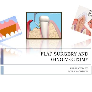
Flap Surgery And Gingivectomy 1g665q
March 2021 0
Gingivectomy 3k581v
December 2019 45
Flap Design For Minor Oral Surgery s12p
October 2019 54
Inscision And Flap Design 4m6u5w
November 2021 0
Flap Procedures 4a4b3c
December 2019 32
Gurney Flap 3n5063
August 2021 0More Documents from "Puneet Sachdeva" 182a3b
Flap Surgery And Gingivectomy 1g665q
March 2021 0
Astrology Reveals The Perfect Girlfriend For Each Zodiac Sign 2t286n
November 2019 80
Anka Shastra Ancient Indian Numerology Jai Guru Dev 2u244z
December 2019 282
Industrial Instrumentation Syllabus 5p6uv
November 2022 0
Migrator For Notes To Sharepoint Pre Migration Planning Guide 642 6v5s28
December 2021 0